Environmental contamination and male reproductive health: (ir) reversible effects in child- and adulthood
Abstract
Infertility affects 10-15% of reproductive-age couples, with causes ranging from genetic factors to unidentified reasons. Environmental conditions, particularly pollutants, play a significant role in male fertility. Yet, public health policies often overlook reproductive health, despite mounting evidence of pollutants’ detrimental repercussion. Understanding this impact is crucial to prevent the effects of dangerous exposure, especially given the high levels of environmental pollutants in today’s world. Most of the previous research about the adverse effects from contaminants has been conducted in rodents, with limited human epidemiological research. This article reviews the evidence about the impact of various contaminants (air pollutants, water contaminants, pesticides, herbicides, radiation, heavy metals, and plastics) on male reproductive health, particularly sperm quality and fertility. The literature suggests that exposure to contaminants during fetal development and childhood has irreversible effects, while those of adult exposure are often reversible. These findings highlight the need to alert society about reproductive health threats from certain contaminants. Public authorities should consider this situation when designing health plans, and individuals envisaging fatherhood should be aware of these risks.
Key words
air pollution; male infertility; pesticides; plastics; radiation; water contaminants
INTRODUCTION
Searching for the term “male infertility” on specialized websites such as Science Direct or PubMed retrieves more than 75,000 papers published in the last ten years. Most of these studies suggest that male infertility may result from decreased seminal quality (Blay et al. 2020), which is assessed based on sperm kinetics, concentration, morphology, and the functionality and integrity of organelles. In our globalized world, environmental pollution is a daily reality for humans from conception onward. When assessing the toxicity of environmental contaminants, their effects on lethality, survival, development, and growth are typically evaluated, but the potential impact on reproduction is often overlooked. Nevertheless, many studies have identified environmental pollution as a factor contributing to male infertility (Fuller et al. 2022). Previous research has consistently demonstrated that heavy metals, microplastics, herbicides, fungicides, and air toxins harm reproductive function. Despite this, whether this damage is irreversible remains unresolved (Blay et al. 2020, Fuller et al. 2022).
While it is now widely accepted that environmental contaminants are pervasive, actions to prevent their harmful impact remain limited (Fuller et al. 2022). To guide future research, questions such as whether these adverse effects can be reversed and how prolonged exposure must be avoided to achieve recovery remain unanswered. Additionally, most studies on the damage caused by nanomolecules and microplastics have been conducted in rodents, and epidemiological data in humans primarily cover adulthood. Thus, there is a critical research gap in understanding how current exposure to environmental pollutants may affect the future reproductive potential of infants.
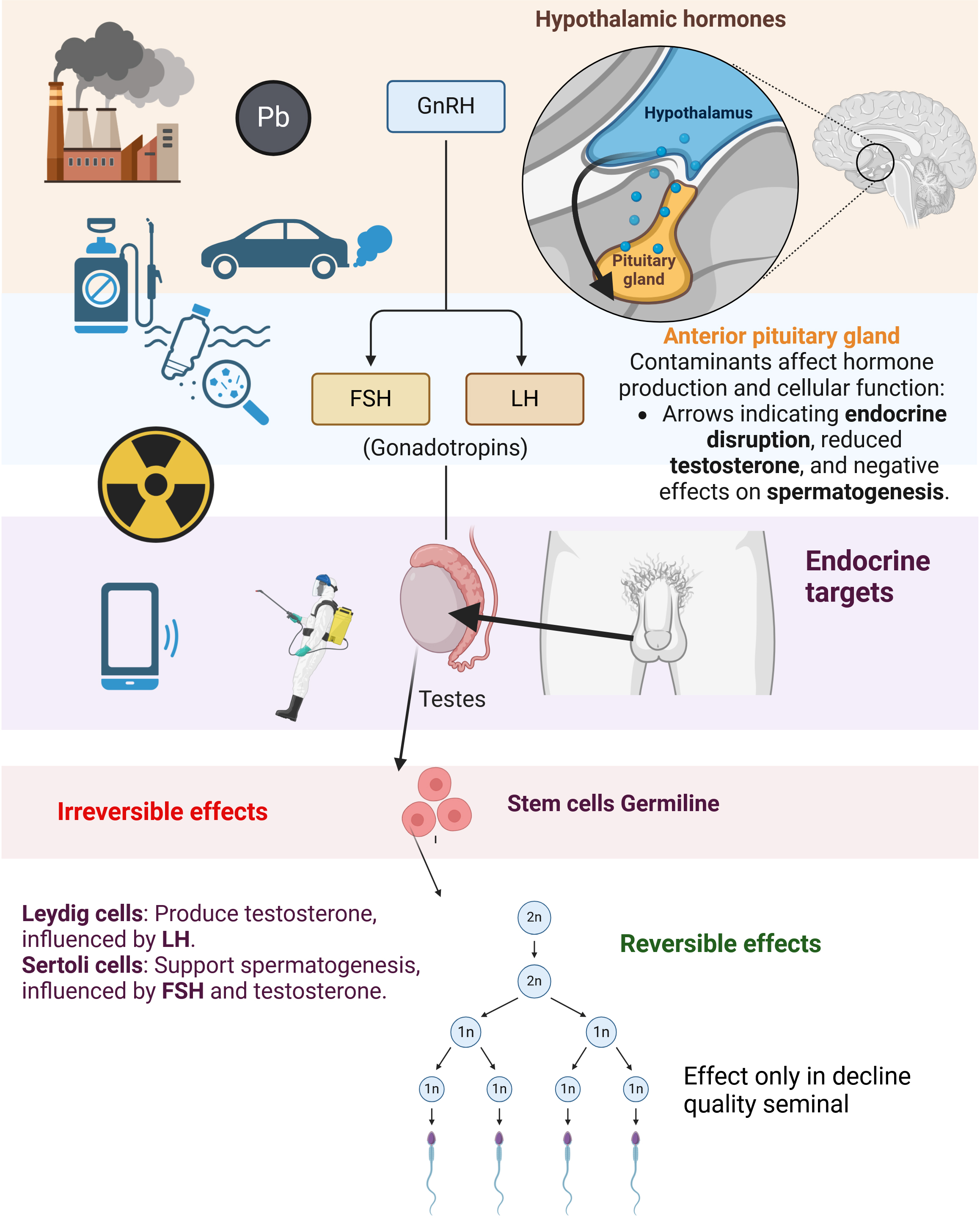
In this review, broad categories of environmental contaminants - such as air pollution, pesticides, plastics, and radiation (Figure 1) - were selected based on their prevalence, scientific relevance, and the documented impacts on reproductive health. Figure 1 illustrates the pathways through which environmental contaminants impact the male reproductive system via the hypothalamic-pituitary-testicular (HPT) axis. Key contaminants—such as heavy metals, microplastics, and pesticides—disrupt endocrine signaling, interfering with hormonal regulation and leading to reduced testosterone production and impaired spermatogenesis. These adverse effects are categorized as either reversible or irreversible based on their persistence and impact on cellular structures.

Reversible and irreversible effects of environmental pollutants on the male reproductive system, including the impact on hormone levels.
Irreversible effects, highlighted in red, primarily target germline stem cells, causing enduring damage that remains even after the cessation of exposure. Such damage compromises long-term spermatogenesis recovery and may lead to permanent infertility. Conversely, reversible effects, marked in green, mainly affect Leydig and Sertoli cells, resulting in a temporary decline in seminal quality. This impairment typically improves upon removal of the contaminants, allowing for the restoration of testicular function and sperm production. These categories represent contaminants with substantial evidence for reproductive toxicity and are among the most widely studied in the context of male infertility. The selection was also driven by the aim to include contaminants with the potential for both short-term and long-term effects on the reproductive system. While there is a plethora of other contaminants not included, these choices reflect the most pressing concerns in the literature and the global exposure patterns that demand further investigation. In addition, this review considers the distinction between reversible and irreversible effects of these contaminants, a crucial factor in understanding their long-term impact. The influence of maternal exposure is also examined, as it can significantly affect fetal development and potentially lead to lifelong reproductive issues. Moreover, the review explores the epigenetic effects of environmental exposure, focusing on how these contaminants may cause heritable changes in gene expression that can affect not only individual health but also future generations.
In view of these considerations, this narrative review aims to compile and critically evaluate the current evidence on the detrimental effects of various environmental factors - such as air pollution, pesticides, plastics, and radiation - on reproductive health. Furthermore, it seeks to investigate whether these adverse effects are reversible or if interventions can mitigate the long-term reproductive damage caused by such exposures.
AIR POLLUTANTS
When addressing the consequences of air pollution, it is important to consider that during certain seasons, such as summer, pollutants dissipate more and are present at lower concentrations in the atmosphere. Previous research has shown that human sperm exposed to high levels of air pollution (particulate matter <10 μm in aerodynamic diameter (PM10), total suspended particulates (TSP), SO2, CO, and NOx), especially in winter, exhibit higher levels of DNA fragmentation (%DFI), as evaluated by the Sperm Chromatin Structure Assay (SCSA), without significant changes in semen volume, sperm motility, vigor, or concentration (Evenson et al. 1980, 1991, 1999, 2002, Sun et al. 2020).
Reactive metabolites from polycyclic aromatic hydrocarbons (PAHs) can induce DNA breaks in spermatids and sperm, potentially leading to infertility or contributing to malformations in the offspring due to shortened telomeres or mutations. Additionally, exposure of adult mice to PAH-derived reactive metabolites causes tandem repeat mutations and hypermethylation in spermatogonia B. While somatic cells can repair DNA damage, spermatids and sperm cannot. Despite this, sperm with damaged DNA can still fertilize oocytes, although they give rise to low-quality embryos and are associated with implantation failure and reduced pregnancy rates. According to Somers et al. (2004) and Yauk et al. (2008), the damage to sperm correlates directly with PAH concentration, and high PAH levels can extend the duration of spermatogenesis in mice by approximately 3.5 times (normal period: 21 days; extended period: 74 days). Yet, PAH metabolites do not affect spermatogonial stem cells, meaning that if exposure to these pollutants ceases before the onset of a new spermatogenic cycle, the adverse effects may be reversed (Binková et al. 1995, Somers et al. 2004). The effects of PAH metabolites are, therefore, considered reversible.
Epidemiological studies conducted by Poongothai et al. (2009), and Ikechebelu et al. (2003) reported that in men exposed to high levels of air pollutants, sperm motility decreases, and the proportions of sperm with DNA damage and morphological abnormalities increase. Benzo(a)pyrene (BaP) and carbon black (CB), originating from both natural (e.g., volcanic activity) and anthropogenic sources (e.g., fossil fuel combustion, industrial activities, forest fires), also have detrimental effects on male fertility. BaP metabolites directly influence the testes by inducing reactive oxygen species (ROS) generation and apoptosis in Leydig cells, leading to a reduction in testosterone synthesis and secretion. Testosterone plays a crucial role in spermatogenesis, as it is produced by Leydig cells in response to luteinizing hormone (LH) and diffuses into Sertoli cells to support sperm production. Consequently, BaP acts as an endocrine disruptor, leading to reduced sperm production and male fertility. Another potential explanation for the adverse effects of air pollutants (BaP and CB) on male fertility is the side effects of lung inflammation. Inflammatory factors may alter the permeability of the blood-testis barrier, allowing pollutants to reach the testes (Skovmand et al. 2018) (Table I).
Most previous studies have been conducted on adult rodents, showing significant damage to testicular tissue, including alterations in the seminiferous tubules and reduced germinal epithelium thickness. Notably, the intergenerational effects of these pollutants differ depending on whether exposure occurs during fetal development, where the damage is permanent, or if adults are exposed for only eight weeks, in which case the damage is reversible (Skovmand et al. 2018). Further research is needed to elucidate the mechanisms of air pollutant action in humans and to better characterize their effects on fetal development, the endocrine system, and spermatogenesis.
WATER CONTAMINANTS
In 2018, the Joint Research Center of the European Commission reported that 17-α-ethinylestradiol (EE2) contamination was present in 98.3% of river samples, 1.2% of lake samples, and 0.5% of coastal water samples in Europe (data from Spain, Greece, and Malta were excluded as these countries did not provide the relevant indicators) (Klaic & Jirsa 2022). An example of such contamination comes from hormonal contraceptives, which do not undergo natural degradation or alteration through solar radiation. As a result, estrogens are not entirely removed during sewage treatment and may be transported through water. It has been reported that while oral contraceptives consist of a combination of estrogen (ethinyl estradiol) and synthetic progestin, residual levels of EE2 remain in water even after wastewater treatment (Klaic & Jirsa 2022). The persistence of pharmaceutical products in drinking water supplies poses a public health concern, including risks to reproductive function (Klaic & Jirsa 2022, Ríos-Sossa et al. 2022). In this context, concerns have been raised about whether chronic exposure to these residual hormone levels, even if marginal, contributes to endocrine dysregulation and adversely affects human health (Klaic & Jirsa 2022).
According to Wee et al. (2021), children in Malaysia ingest twice as much EE2 through drinking water as adults on a daily basis. Estradiol is a crucial inhibitor of luteinizing hormone (LH) production and, in males, it limits spermatogenesis and reduces testosterone synthesis by Leydig cells. While exposure to environmental estrogens does not appear to induce permanent changes in the structure and function of adult male reproductive organs, it does lead to reduced sperm motility and concentration, indicating a negative impact on spermatogenesis during exposure (Wee et al. 2021) (Table I).
In rodents, fetal exposure to water contaminants, such as Bisphenol A (prevalent at 66.4 ng/L), does not appear to alter fetal testosterone production, but it does affect the expression of androgen receptor (AR) and estrogen receptor 1 (ESR1), resulting in the underdevelopment of the epididymal duct epithelium and alterations in the prostate and accessory glands (Atanassova et al. 2001). Epidemiological studies have shown that exposure to estrogens during pre- and postnatal stages reduces semen quality; however, most studies focus on sperm and do not evaluate AR and ESR1 expression (Atanassova et al. 2001). Research in humans should, therefore, investigate whether drinking water containing hormone residues affects AR and ESR1 expression. Additionally, while EE2 and Bisphenol A exposure in humans has been reported to decrease seminal quality, the effects on fertility have been insufficiently studied. Notably, studies in rodents have demonstrated that such exposure impairs urogenital system development and fertility and negatively affects secondary sexual characteristics (Ciślak et al. 2023).
Poly- and perfluoroalkyl substances (PFAS), including perfluorooctanoic acid (PFOA) and perfluorooctane sulfonate (PFOS), are present in drinking water at concentrations exceeding 70 ng/L, posing a public health concern. Exposure to PFOS and PFOA increases the percentages of sperm with coiled tails (Specht et al. 2012) and with fragmented DNA, and decreases sperm concentration and motility (Toft et al. 2012). As these agents act as endocrine disruptors, their effects are considered irreversible (Atanassova et al. 2001, Tsai et al. 2015) (Figure 1). Furthermore, high concentrations of PFOA (>9.8 ng/mL) and PFOS (>13.3 ng/mL) are negatively associated with follicle-stimulating hormone (FSH) and testosterone levels in serum, as PFOS and PFOA reduce cholesterol, which is essential for steroid hormone synthesis (Tsai et al. 2015). Given this evidence, the physical, chemical, and biological quality of drinking water, as well as its treatment, remains a critical issue. It is not enough to test for fecal coliforms or heavy metals; hormonal residues, PFOS, and PFOA must also be monitored. Moreover, the effects of long-term exposure to these water contaminants on human fertility warrant further investigation.
PESTICIDES
Humans can be contaminated with pesticides primarily through ingesting contaminated food, direct contact with chemical products during agricultural application, or inhaling airborne particles after spraying. During fetal development, the timing of pesticide exposure is crucial in determining whether damage to reproductive organs and/or infertility can be reversed (Kalb et al. 2016, Vergouwen et al. 1993). Xenoestrogenic (alkylphenols and phytoestrogens) and antiandrogenic pesticides (linuron, procymidone, p,p’-dichlorodiphenyldichloroethylene, fenarimol, and viclozolin metabolites) are known to have significant effects on testicular tissue during fetal and neonatal stages in rodents (de Castro et al. 2018, Kalb et al. 2016, Mikkilä et al. 2006, Vilela et al. 2014, Saalfeld et al. 2019). It is, therefore, evident that exposure to pesticides during the neonatal stage causes damage that manifests in adulthood. Studies in rodents suggest that this injury may be irreversible, as it is directly linked to endocrine-active compounds (Kalb et al. 2016, Mikkilä et al. 2006, Saalfeld et al. 2018, 2019) (Figure 1). Epidemiological studies by Weidner et al. (1998), Garcia-Rodriguez et al. (1996) and Skakkebæk et al. (2001) showed that workers occupationally exposed to pesticides exhibited an increased risk of pathologies such as cryptorchidism, testicular cancer, and penile abnormalities, and their children were more likely to develop genital malformations.
Exposure to Bis-A reduces testicular size by about 30-45% (compared to typical testicular composition), which is associated with a smaller testicular parenchyma and reduced testosterone levels. This is explained by the role of FSH, which interacts with its receptors on Sertoli cells, thereby stimulating spermatogenesis and indirectly affecting testosterone production by Leydig cells (Atanassova et al. 2000). Nevertheless, the effects of Bis-A exposure during embryonic or fetal development on adult male fertility have not yet been studied and should be the focus of future research (Table I).
A previous study found that men exposed to 1,1,1-trichloro-2,2-bis(chlorodiphenyl)ethane (DDT) had impaired sperm quality (De Jager et al. 2006); however, this alteration could be reversed, as the proportion of morphologically normal sperm increased and the percentage of sperm with abnormal heads and tails decreased when exposure was reduced by 25%. It should be noted that only post-pubertal men aged 18 to 49 years were included in this study. A similar pattern was observed in individuals exposed to glyphosate-based herbicides (GBHs), as there was a decrease in progressive sperm motility and viability, along with an increase in sperm DNA damage (Nerozzi et al. 2020) (Table I). Both GBHs and DDT exert endocrine-disruptive effects, as the estrogen increase caused by pesticides reduces testosterone secretion through activating monooxygenase enzymes in the liver and suppresses LH secretion by the pituitary gland. In adult males (i.e., post-puberty), this can interfere with the function of Sertoli cells, which rely on testosterone and LH. Prolonged exposure to these agents ultimately leads to a reduction in sperm production.
An important point to consider in occupational exposure studies to pesticides, such as endosulfan, is the individual variation in contaminant absorption, which can affect the comparability of serum levels among exposed workers (Dalvie et al. 2009). In the study of (Dalvie et al. 2009), baseline serum endosulfan levels were elevated among all participants, indicating significant environmental exposure in rural areas, even before the annual spraying. Both applicators (75%) and non-applicators (>50%) showed substantial increases in serum endosulfan levels, demonstrating that exposure to the pesticide occurs not only through direct contact but also via spray drift. Age was identified as the only significant confounding factor in predicting post-spraying increases, suggesting that individual factors such as metabolism and absorption capacity must be considered when interpreting occupational exposure results. These findings highlight the importance of assessing individual variation to obtain a more accurate understanding of the risks associated with pesticide exposure. The study by Chetty-Mhlanga et al. (2018) was a longitudinal investigation about how pesticide exposure (organophosphate metabolites, dialkyl phosphates (DAPs), and pyrethroids) affects the reproductive health and neurobehavioral development of children aged 9 to 16 in agricultural areas. Using a combination of biomonitoring techniques, such as urine and hair samples, along with environmental assessments of air and water, the study provided a comprehensive evaluation of the effects of pesticide exposure on reproductive hormone levels and sexual maturity. The research included children at various stages of development, allowing for the analysis of pubertal and neurobehavioral changes over time. By incorporating data on co-exposures, such as media use and alcohol consumption, the study offered a detailed understanding of the factors influencing exposure and their effects. The results could be helpful to both local agricultural communities, providing evidence-based recommendations for health promotion and pesticide regulation. This was the first longitudinal study to examine the impact of contemporary agricultural pesticides on children’s reproductive health, addressing gaps and inconsistencies in previous research on neurobehavioral outcomes. Based on all the evidence mentioned above, one can conclude that pesticides cause irreversible and permanent damage to reproductive function when exposure occurs during the perinatal period, resulting in reduced testicular size and decreased sperm production (Kalb et al. 2016). If exposure occurs after puberty, the damage may be reversible, as lowering the substance by more than 25% results in decreased sperm damage.
HERBICIDES
Due to its selective control of grasses and weeds, atrazine (ATR, (2-chloro-4-ethylamino-6-isopropylamino-1,3,5-triazine)) is one of the most widely used herbicides in agriculture (García et al. 2012). Yet, several studies reported the behavior of ATR as an endocrine disruptor in mammals. The mechanism by which ATR disrupts the endocrine system is related to the binding and further inhibition of the enzyme phosphodiesterase (PDE). Inhibition of PDE increases the levels of cyclic adenosine monophosphate (cAMP), which upregulates the expression of the CYP19 gene that encodes for aromatase enzyme (Sanderson et al. 2000). The function of aromatase is to convert testosterone into estrogen, the final effect produced by ATR thus being an elevation of estrogen levels in cells (Sanderson et al. 2000).
In adult rodents (120 days old), exposure to ATR adversely affects the reproductive system, as it reduces the weight of testicles; increases the size of the adrenal gland; induces the dilation and disorganization of the seminiferous tubules; causes irregularities in the nucleus of Leydig cells; and disrupts the cell junctions between Sertoli cells and germ cells, thereby decreasing testosterone levels (Song et al. 2014). Besides, ATP production in ejaculated sperm is highly affected by direct exposure of rat males to ATR. Specifically, ATR inhibits ATP synthase, which in turn affects other mitochondrial functions. This results in decreased sperm viability and, particularly, motility (Abarikwu et al. 2010, 2015).
On the other hand, exposure to ATR during gestation and lactation also causes reproductive damage to pups. In rats, exposure of pregnant females to ATR during early development and the gestational period decreases the weight of testes, epididymes, and the entire body of pups (Song et al. 2014); reduces the anogenital distance; and decreases the expression of stereidogenic and androgen genes, which has toxic effects for the fetus across the male lineage in F1 and F2 generations (Pandey et al. 2021).
In the case of humans, exposure to ATR decreases sperm motility (Hase et al. 2008), and alters the composition of seminal plasma (Rodríguez-Robledo et al. 2022), which has a major impact on male fertility. This supports the need of testing the effects of toxicants not only in sperm but also in seminal plasma, even employing techniques such as chromatography. In humans, changes in seminal plasma as consequence of exposure to environmental toxicants could have a detrimental impact on sperm physiology (sperm motility) and could affect the role played by seminal plasma in the female reproductive tract. Remarkably, the sole evaluation of sperm quality without envisaging the potential role of seminal plasma often shifts the focus to the woman when a couple has problems of fertility; this approach, however, forgets about the potential role of seminal plasma in the whole physiological process of fertilization and embryo implantation (Rodríguez-Robledo et al. 2022).
Because evidence regarding the irreversible effects of prenatal exposure to herbicides in rodents accumulates, further studies should determine if this is also the case in humans. In addition, while the impact of ATR on the sperm quality of human adults appears to be transient, this does not exclude that the repercussion observed in the seminal plasma could last longer. For this reason, future investigations should also be aimed to address if the seminal plasma from males exposed to ATR exerts a disadvantageous effect on the reproductive success and embryo development.
MICROPLASTICS AND PLASTICIZERS
It is estimated that about 800 million tons of plastic end up in the oceans every year (Li et al. 2021, Steer et al. 2017). As the industry grows and habits do not change, researchers estimate that the amount of plastic waste will exceed about 11 billion tons by 2050. In recent years, consensus has been reached about the problem being neglected by public health policies. Plastics undergo photo (ultraviolet light) and biodegradation (wave motion, temperature, among others), thus resulting in microplastics (diameter less than 5 mm) (Steer et al. 2017). Humans are contaminated with microplastics through ingesting contaminated food and water, inhaling airborne particles, and consuming seafood, as microplastics are present in the oceans and eaten by marine organisms. Additionally, everyday products like cosmetics and synthetic clothing can release microplastics, which are eventually absorbed by the body. The research on this topic is still in its infancy, as most previous studies were conducted in rodents rather than in humans. Such investigations in rodents only examined the effects of exposing post-pubertal animals to microplastics, and observed a decrease in sperm motility and sperm concentration, an increase in the proportions of morphologically abnormal sperm and sperm with DNA damage, and a narrowing of the diameter of seminiferous tubules (Ijaz et al. 2022, Jin et al. 2021, Li et al. 2021). Since no examinations during gestations have hitherto been carried out, whether the injuries induced by microplastic pollutants are reversible or irreversible remains unknown. While no study has looked into the effects on spermatogonia, it has been estimated that the dose of microplastics that human beings may be exposed to (16 μg/kg/day) can be detrimental to sperm quality (Ijaz et al. 2022).
Bisphenol A (BPA) is a synthetic organic compound widely used to produce plastics (toys, bottles, flooring materials, food packaging and many others). Previous studies in rodents proved that exposure to BPA, during either pre or post-natal stages, decreases the size of testicles, epididymes and preputial glands, which results in reduced seminal quality. Noticeably, these effects are persistent in the offspring until adulthood. Also, in rodents, BPA was revealed to inhibit the expression of StAR and 17-β-hydroxysteroid dehydrogenase (17β-HSD), which leads to a decline in the secretion of androgens by Leydig cells. In humans, epidemiological studies (Braun et al. 2012, Mínguez-Alarcón et al. 2018) revealed that not only was BPA detected in the urine of women exposed to this toxicant during pregnancy (first, second, or third trimester), but its concentration in serum was also higher. Exposure to BPA during gestation and lactation has, therefore, harmful, irreversible effects on the physiology of conceptus, as it impairs the development of reproductive organs, their response to hormonal stimuli, and their ability to produce sperm. Remarkably, a matter of concern is that such damage can be passed on to the next generations, thus compromising the development of the reproductive system in the offspring and causing male infertility; for this reason, the effects on epigenetic signatures should also be considered in future studies. Because of all the aforementioned, BPA is no longer included as a plastic component, as if it came into contact with children during the developmental stages, it would have a very negative repercussion (Al-Saleh et al. 2019, Braun et al. 2012, Caporossi et al. 2020, Harchegani et al. 2019, Mínguez-Alarcón et al. 2018, Thurston et al. 2016). In human adults, exposure to BPA inhibits the synthesis of FSH, increases the plasma levels of LH and decreases those of testosterone and prolactin. This increase in plasma LH is related to the negative feedback caused by the decrease in testosterone concentration, which interferes with sperm production. Thus, in adults, BPA appears to decrease sperm concentration, motility, and the proportions of morphologically normal sperm. It should be emphasized that in adults where the development of the reproductive system has already been completed, the deleterious effects on seminal quality are potentially reversible when exposure ceases (Caporossi et al. 2020) (Table I, Figure 1).
Diethylhexyl phthalate (DEHP) is a synthetic chemical widely used in the fabrication of medical devices, toys, and food packages, among others. The forms of contamination in humans are via ingestion, inhalation, and topical absorption. Exposure to phthalate during the neonatal period reduces the anogenital distance in boys (Swan et al. 2005). The effect of DEHP would be related to its antiandrogenic nature (Curi et al. 2019, Hutson et al. 2013, Sunman et al. 2019), as the anogenital distance is known to be associated with the development of the male reproductive system, which is driven by testosterone levels. This decrease in the anogenital distance impacts the adult life of men, and is linked to reduced sperm concentration. While exposure to phthalates has been found to cause pathologies such as cryptorchidism and hypospadias in rodents, prenatal exposure to these toxicants has not been reported to exert such a detrimental impact (Hutson et al. 2013, Sunman et al. 2019); notwithstanding, whether concentrations greater than those tested could be a risk factor still needs to be elucidated. Increased levels of DEPH in urine are associated with high FSH (β = 0.118) and LH concentrations (β = 0.099) in men, thus indicating abnormal sperm production; and low testosterone (β = −0.086) and estrogen levels, which indicates aromatase inhibition (Al-Saleh et al. 2019). These decreased testosterone levels after DEPH exposure reflect a disruption in the regulation of the hypothalamic-pituitary-thyroid (HPT) axis, and the higher production of FSH and LH produces an imbalance because Sertoli and Leydig cells do not have enough receptors for FSH and LH, which alters spermatogenesis (Al-Saleh et al. 2019). Consequently, adults exposed to DEPH show low sperm concentration and few cells with normal morphology, due to endocrine dysregulation (Al-Saleh et al. 2019). In rodents, if the exposure to 100 mg/Kg BW/day of DEPH is shorter than 42 days (i.e., the period of two spermatogenic waves), the effects on seminal quality may be reversible; a longer period, however, causes irreversible damage (Al-Saleh et al. 2019, Caporossi et al. 2020, Harchegani et al. 2019, Thurston et al. 2016) (Figure 1). In humans, a previous work demonstrated that exposure to DEHP reduced sperm concentration by 29% (Mínguez-Alarcón et al. 2018).
Plasticizers, liquids added to microplastic or nanoplastics (defined as particles smaller than 5 mm or ranging between 1 and 100 nm in size) have different mechanisms of action in organisms. The most remarkable issue, however, is that any exposure before puberty (childhood) causes irreversible damage to the male reproductive system. Conversely, while exposure during adulthood may reduce seminal quality, the adverse effect relies on the dose and period of exposure and appears to be reversible (Ijaz et al. 2022, Jin et al. 2021, Li et al. 2021).
RADIATIONS
Humans are inevitably exposed to radiations, which can be either non-ionizing or ionizing. According to Kesari et al. (2018), non-ionizing radiations consist of low-frequency radiation (UV), electromagnetic radiation and radio frequencies. Remarkably, humans are continuously exposed to non-ionizing electromagnetic radiation of low intensity for long periods through cell phones, antennas, light bulbs, and radars. Ionizing radiations are those that have enough energy to remove electrons from atoms, producing electromagnetic waves, α and β particles. Spermatogonia are radiosensitive and can die in response to ionizing radiation. Other testicular, further differentiated germ cells such as spermatocytes and spermatids are even more sensitive to ionizing radiation. For this reason, the testis is considered the most radiosensitive to ionizing radiation organ in the human body (Xu et al. 2008). Exposure to electromagnetic radiation causes a reduction in serum testosterone levels, which leads to a decrease in sperm motility and concentration (D’Autreaux & Toledano 2007, De Iuliis et al. 2009, Forgács et al. 2006, Willians & Fletcher 2010). It has also been reported that keeping the cell phone in the pocket of men (10 cm from the testicles) interferes with seminal quality, impairing sperm motility and viability (D’Autreaux & Toledano 2007). In mice, Forgács et al. (2006) identified that seminiferous tubules became narrower after exposure to an electromagnetic wave of 1800 MHz from a cell phone having a specific absorption rate (SAR) of 0.023 W/Kg. This electromagnetic radiation also reduced testosterone production, possibly affecting spermatogenesis (Table I, Figure 1).
Exposure to an ionizing radiation dose of 0.3 grays (Gy; 30 rad) during embryonic development can be teratogenic, mutagenic, or carcinogenic. The fetus is more susceptible to damage during the period from two to 15 weeks post-conception (organogenesis), and previous studies conducted in women showed that this is the most critical phase for radiation-induced miscarriage (Willians & Fletcher 2010). Wistar rats exposed to non-ionizing radiation (frequency of 60 HZ and magnetic intensity of 1 mT) for 21 days of gestation were found to undergo a significant reduction in the diameter of their seminiferous tubules, without changes in the other histological parameters of the testis, epididymis, seminal vesicles and the prostate (Çelik et al. 2012, Tenorio et al. 2012). Another study found that exposure to electromagnetic radiation over the gestation of adult males may lead to subfertility and infertility because of testicular degeneration, increased apoptosis rate in the seminiferous epithelium, vacuolization of the cytoplasm of Sertoli cells, and decreased sperm viability and motility, without altering testosterone synthesis (Sharma et al. 2021). In addition, testicles are very sensitive to thermal variations, so the increment of temperature caused by ionizing radiation interferes with spermatogenesis negatively (Xu et al. 2008). On the other hand, whilst exposure of men to a low dose of 0.023 W/Kg radiation for long periods leads to a reduction in sperm count and motility, and also narrows the seminiferous tubule diameter, a short period of exposure has no effect. Further studies are thus needed to clarify how electromagnetic radiation acts on the human testicular tissue, including under which conditions (type of radiation, time of exposure, frequency, and male age) adverse effects are irreversible.
HEAVY METALS
Metals are natural components of the environment, most of which are essential micronutrients for living organisms. They can be found in terrestrial and aquatic systems as well as in the air. They can concentrate through food webs, from which species at the top of the food chain can accumulate high levels of metals (Hernandez et al. 1999). Unlike many organic compounds, metals cannot be easily metabolized into less toxic compounds, so they have long residence times in soil and might cause harmful effects long after the pollution by the metal itself has occurred (Berglund et al. 2009).
Bergamo et al. (2016) found a large amount of Zn, Cr and Cu in the ejaculates of men living in areas with high environmental impact. Previous research supports that heavy metals disrupt the hypothalamic-pituitary-testis axis, as they interfere with androgen production in two possible ways: either through receptor alteration or by directly affecting gene transcription (De Angelis et al. 2017, Pavlova & Atanassova 2018). Pb, Cd and As have recently been identified as the main toxicants altering the function of reproductive organs, such as the prostate, increasing the percentages of sperm with high DNA fragmentation and low degree of chromatin condensation, and reducing the ejaculate volume (De Angelis et al. 2017, Pavlova & Atanassova 2018). According to the World Health Organization (WHO), while there are no safe exposure levels for Pb, those ≥ 10 μg/dL harm the male reproductive system (Pavlova & Atanassova 2018). Indeed, the presence of 0.5 μg/L Pb in men’s seminal plasma is associated with spontaneous abortions due to sperm DNA damage (Pavlova & Atanassova 2018). Previous research in rodents reported that the administration of antioxidants, such as ascorbic acid combined with thiamine for six weeks (two periods of spermatogenesis) partially neutralizes the detrimental action of Pb on the reproductive system (Wang et al. 2006) (Table I).
Bioaccumulation and half-life (between 20 and 40 years) of cadmium (Cd) make it an important metal in reproductive toxicology. Noticeably, Cd concentration in seminal plasma is associated with reduced sperm motility and concentration, and increased sperm DNA damage and oxidative stress (Mendiola et al. 2011, Pant et al. 2015). How Cd precisely works in humans is unknown, but studies in rodents showed that it acts as an endocrine disruptor (Pavlova & Atanassova 2018) (Table I).
Finally, in humans, Mirnamniha et al. (2019) demonstrated that Selenium (Se) can act by partially neutralizing the harmful effects of Cd and Pb. Se is an essential constituent of the enzyme glutathione peroxidase, which plays a crucial role in the antioxidant defense scavenging reactive oxygen metabolites. Besides being an excellent antioxidant, it prevents the damaging effects caused by oxidative stress after a treatment period of three months. As all studies conducted in humans involved adults, the impact of Se on children is not known.
CONCLUSIONS
The causes underlying infertility in men have been investigated in the last years, with accumulating evidence now supporting that environmental factors can also alter sperm quality, function, and fertility. It is known that these environmental effects do not happen alone; they can be amplified by lifestyle, genetic factors, and especially the forms of contamination and combination of these reagents.
In the case of adults, the effects of most environmental contaminants appear to be reversible, as sperm quality can be partially restored when individuals are removed from exposure. One, however, must notice that most studies focus on conventional spermiogram parameters (i.e., motility, concentration, and morphology) but do not address whether traces of contaminants are present in seminal plasma. Yet, and as illustrated in the current review, the presence of contaminants in seminal plasma can be associated with reduced sperm quality and fertility. In addition, the assessment of other sperm function variables may be required to understand the actual impact of exposing men to contaminants. Unfortunately, the exact mechanisms through which these contaminants affect reproductive function remain, in most cases, unknown, and should be investigated in the future.
Another conclusion of this review is that exposure to environmental contaminants during fetal development and childhood may impact reproductive function until the second generation. For some compounds, only literature on rodents is available and there is a lack of epidemiological data on humans. Further research is, therefore, warranted to address how all these environmental pollutants compromise reproductive function in children and during fetal development.
ACKNOWLEDGMENTS
The authors acknowledge the support from the Regional Government of Catalonia, Spain (Grant: 2021-SGR-00900) and the Catalan Institution of Research and Advanced Studies (ICREA). C.D. Corcini (310003/2021-0 and 401592/2022-7) and A.S. Varela Junior (307678/2022-9) are research fellow from the Conselho Nacional de Desenvolvimento Científico e Tecnológico.
REFERENCES
- ABARIKWU SO, ADESIYAN AC, OYELOJA TO, OYEYEMI MO & FAROMBI EO. 2010. Changes in Sperm characteristics and Induction of Oxidative Stress in the Testis and Epididymis of Experimental Rats by a Herbicide, Atrazine. Arch Environ Contam Toxicol 58: 874-882. doi: 10.1007/s00244-009-9371-2.
- ABARIKWU SO, AKIRI OF, DUROJAIYE MA & ADENIKE A. 2015. Combined effects of repeated administration of Bretmont Wipeout (glyphosate) and Ultrazin (atrazine) on testosterone, oxidative stress and sperm quality of Wistar rats. Toxicol Mech Methods 25: 70-80. doi: 10.3109/15376516.2014.989349.
- AL-SALEH I, COSKUN S, AL-DOUSH I, AL-RAJUDI T, ABDULJABBAR M, AL-ROUQI R, PALAWAN H & AL-HASSAN S. 2019. The relationships between urinary phthalate metabolites, reproductive hormones and semen parameters in men attending in vitro fertilization clinic. Sci Total Environ 658: 982-995. doi: 10.1016/j.scitotenv.2018.12.261.
- ATANASSOVA N, MCKINNELL C, TURNER KJ, WALKER M, FISHER JS, MORLEY M, MILLAR MR, GROOME NP & SHARPE RM. 2000. Comparative effects of neonatal exposure of male rats to potent and weak (environmental) estrogens on spermatogenesis at puberty and the relationship to adult testis size and fertility: evidence for stimulatory effects of low estrogen levels. Endocrinology 141: 3898-907. doi:10.1210/endo.141.10.7723.
- ATANASSOVA N, MCKINNELL C, WILLIAMS K, TURNER KJ, FISHER JS, SAUNDERS PT, MILLAR MR & SHARPE RM. 2001. Age-, cell- and region-specific immunoexpression of estrogen receptor alpha (but not estrogen receptor beta) during postnatal development of the epididymis and vas deferens of the rat and disruption of this pattern by neonatal treatment with diethylstilbestrol. Endocrinology. 142: 874-886. doi: 10.1210/endo.142.2.7978.
- BERGAMO P, VOLPE MG, LORENZETTI S, MANTOVANI A, NOTARI T, COCCA E, CERULLO S, DI STASIO M, CERINO P & MONTANO L. 2016. Human semen as an early, sensitive biomarker of highly polluted living environment in healthy men: A pilot biomonitoring study on trace elements in blood and semen and their relationship with sperm quality and RedOx status. Reprod Toxicol 66: 1-9. doi:10.1016/j.reprotox.2016.07.018.
- BERGLUND MMÅ, KLAMINDER J & NYHOLM NEI. 2009. Effects of reduced lead deposition on pied flycatcher (Ficedulahypoleuca) nestlings: tracing exposure routes using stable lead isotopes. Environ Sci Technol 43: 208-213. doi:10.1021/es801723q.
- BINKOVÁ B, LEWTAS J, MISKOVÁ I, LENÍCEK J & SRÁM R. 1995. DNA adducts and personal air monitoring of carcinogenic polycyclic aromatic hydrocarbons in an environmentally exposed population. Carcinogenesis 16: 1037-46. doi: 10.1093/carcin/16.5.1037.
- BLAY RM, PINAMANG AD, SAGOE AE, OWUSU EDA, KONEY NK-K & ARKO-BOHAM B. 2020. Influence of Lifestyle and Environmental Factors on Semen Quality in Ghanaian Men. Int J Reprod Med 2020: 6908458. doi: 10.1155/2020/6908458.
- BRAUN JM, SMITH KW, WILLIAMS PL, CALAFAT AM, BERRY K, EHRLICH S & HAUSER DR. 2012. Variability of urinary phthalate metabolite and bisphenol A concentration before and during pregnancy. Environ Health Perspect 120: 739-74. doi:10.1289/ehp.1104139.
- CAPOROSSI L, ALTERI A, CAMPO G, PACI E, TRANFO G, CAPANNA S, PAPALEO E, PIGINI D, VIGANÒ P & PAPALEO B. 2020. Cross Sectional Study on Exposure to BPA and Phthalates and Semen Parameters in Men Attending a Fertility Center. Int J Environ Res Public Health 17: 489. doi:10.3390/ijerph17020489.
- ÇELIK S, ARIDOGAN IA, IZOL V, ERDOĞAN S, POLAT S & DORAN S. 2012. An evaluation of the effects of long-term cell phone use on the testes via light and electron microscope analysis. Urol 79: 346-350. doi:10.1016/j.urology.2011.10.054.
- CHETTY-MHLANGA S, BASERA W, FUHRIMANN S, PROBST-HENSCH N, DELPORT S, MUGARI M, WYK JV, RÖÖSLI M & DALVIE MA. 2018. A prospective cohort study of school-going children investigating reproductive and neurobehavioral health effects due to environmental pesticide exposure in the Western Cape, South Africa: study protocol. BMC Pub Health 18: 857. doi.org/10.1186/s12889-018-5783-0.
- CIŚLAK M, KRUSZELNICKA I, ZEMBRZUSKA J & GINTER-KRAMARCZYK D. 2023. Estrogen Pollution of the European Aquatic Environment: A Critical Review. Water Res 229: 119413. doi: 10.1016/j.watres.2022.119413.
- CURI TZ, DA SILVA GN, PASSONI MT, TOLOUEI SEL, MELDOLA H, ROMANO RM, GRECHI N, DALSENTER PR & MARTINO-ANDRADE AJ. 2019. In Utero and lactational exposure to diisopentyl phthalate induces fetal toxicity and antiandrogenic effects in rats. Toxicol Sci 171(2): 347-358. doi:10.1093/toxsci/kfz159.
- D’AUTREAUX B & TOLEDANO MB. 2007. ROS as signalling molecules: Mechanisms that generate specificity in ROS homeostasis. Nat Rev Mol Cell Biol 8: 813-824. doi:10.1038/nrm2256.
- DALVIE MA, AFRICA A, SOLOMONS A, LONDON L, BROUWER D & KROMHOUT H. 2009. Pesticide exposure and blood endosulfan levels after first season spray amongst farm workers in the Western Cape, South Africa. J Environ Sci Health 44(Part B): 271-277. doi:10.1080/03601230902728351.
- DE ANGELIS C, GALDIERO M, PIVONELLO C, SALZANO C, GIANFRILLI D, PISCITELLI P, LENZI A, COLAO A & PIVONELLO R. 2017. The environment and male reproduction: The effect of cadmium exposure on reproductive function and its implication in fertility. Reprod Toxicol 73: 105-127. doi:10.1016/j.reprotox.2017.07.021.
- DE CASTRO TF, SAALFELD GQ, VARELA JUNIOR AS, PADILHA FF, SANTOS KS, PIRES DM, PEREIRA JR, CORCINI CD & COLARES EP. 2018. Triphenyltin exposition induces spermatic parameter alters of Calomys laucha species. Chemosphere 211: 1176-82. doi: 10.1016/j.chemosphere.2018.08.048.
- DE IULIIS GN, NEWEY RJ, KING BV & AITKEN RJ. 2009. Mobile Phone Radiation Induces Reactive Oxygen Species Production and DNA Damage in Human Spermatozoa in Vitro. PLoS ONE 4(7): e6446. doi:10.1371/journal.pone.0006446.
- DE JAGER C, FARIAS P, BARRAZA-VILLARREAL A, AVILA MH, AYOTTE P, DEWAILLY E, DOMBROWSKI C, ROUSSEAU F, SANCHEZ VD & BAILEY JL. 2006. Reduced Seminal Parameters Associated With Environmental DDT Exposure and p,p’-DDE Concentrations in Men in Chiapas, Mexico: A Cross-Sectional Study. J Androl 27: 16-27. doi: 10.2164/jandrol.05121.
- EVENSON DP, DARZYNKIEWICZ Z & MELAMED MR. 1980. Relation of mammalian sperm chromatin heterogeneity to fertility. Science 210: 1131-1133. doi: 10.1126/science.7444440.
- EVENSON DP, JOST LK, BAER RK, TURNER TW & SCHRADER SM. 1991. Individuality of DNA denaturation patterns in human sperm as measured by the sperm chromatin structure assay. Reprod Toxicol 5: 115-125. doi: 10.1016/0890-6238(91)90039-i.
- EVENSON DP, JOST LK, MARSHALL D, ZINAMAN MJ, CLEGG E, PURVIS K, DE ANGELIS P & CLAUSSEN OP. 1999. Utility of the sperm chromatin structure assay as a diagnostic and prognostic tool in the human fertility clinic. Hum Reprod 14: 1039-1049. doi: 10.1093/humrep/14.4.1039.
- EVENSON DP, LARSON KL & JOST LK. 2002. Sperm chromatin structure assay: its clinical use for detecting sperm DNA fragmentation in male infertility and comparisons with other techniques. J Androl 23: 25-43. doi:10.1002/j.1939-4640.2002.tb02599.x.
- FORGÁCS Z, SOMOSY Z, KUBINYI G, BAKOS J, HUDÁK A, SURJÁN A & THURÓCZY G. 2006. Effect of whole-body 1800MHz GSM-like microwave exposure on testicular steroidogenesis and histology in mice. Reprod Toxicol 22: 111-117. doi:10.1016/j.reprotox.2005.12.003.
- FULLER R ET AL. 2022. Pollution and health: a progress update. Lancet Planet Health 6: e535-547. doi:10.1016/S2542-5196(22)00090-0.
- GARCÍA MÁ, SANTAEUFEMIA M & MELGAR MJ. 2012. Triazine residues in raw milk and infant formulasfrom spanish northwest, by a diphasic dialysis extraction. Food Chem Toxicol 50(3-4): 503-510. doi: 10.1016/j.fct.2011.11.019.
- GARCIA-RODRIGUEZ J, GARCÍA-MARTÍN M, NOGUERAS-OCAÑA M, LUNA-DEL-CASTILLO JDDIOS, ESPIGARES GARCÍA M, OLEA N & LARDELLI-CLARET P. 1996. Exposure to pesticides and cryptorchidism: geographical evidence of a possible association. Environ Health Perspect 104: 1090-1095. doi: 10.1289/ehp.104-1469503.
- HARCHEGANI AB, RAHMANI H, TAHMASBPOUR E & SHAHRIARY A. 2019. Hyperviscous semen causes poor sperm quality and male infertility through induction of oxidative stress. Curr Urol 13(1): 1-6. doi: 10.1159/000499302.
- HASE Y, TATSUNO M, NISHI T, KATAOKA K, KABE Y, YAMAGUCHI Y, OZAWA N, NATORI M, HANDA H & WATANABE H. 2008. Atrazine binds to F1F0ATP synthase and inhibits mitochondrial function in sperm. Biochem Biophys Res Commun 366: 66-72. doi: 10.1016/j.bbrc.2007.11.107.
- HERNANDEZ ML ET AL. 1999. Accumulation of heavy metals and As in wetland birds in the area around Doñana National Park affected by the Aznalcollar toxic spill. Sci Total Environ 242: 293-308. doi: 10.1016/s0048-9697(99)00397-6.
- HUTSON JM, SOUTHWELL BR, LI R, LIE G, ISMAIL K, HARISIS G & CHEN N. 2013. The regulation of testicular descent and the effects of cryptorchidism. Endocr Rev 34(5): 725-752. doi: 10.1210/er.2012-1089.
- IJAZ MU, AYAZ F, MUSTAFA S, ASHRAF A, ALBESHR MF, RIAZ MN & MAHBOOB S. 2022. Toxic effect of polyethylene microplastic on testicles and ameliorative effect of luteolin in adult rats: Environmental challenge. J King Saud Univ - Sci 34: 102064. doi: 10.1016/j.jksus.2022.102064.
- IKECHEBELU JI, ADINMA JIB, ORIE EF & IKEGWUONU SO. 2003. High Prevalence of Male Infertility in Southeastern Nigeria. J Obstet Gynaecol 23: 657-659. doi: 10.1080/01443610310001604475.
- JIN H, MA T, SHA X, LIU Z, ZHOU Y, MENG X, CHEN Y, HAN X & DING J. 2021. Polystyrene microplastics induced male reproductive toxicity in mice. J Hazard Mater 401: 123430. doi: 10.1016/j.jhazmat.2020.123430.
- KALB AC, KALB AL, CARDOSO TF, FERNANDES CG, CORCINI CD, VARELA JUNIOR AS & MARTINEZ PE. 2016. Maternal Transfer of Bisphenol a during nursing causes sperm impairment in male offspring. Arch Environ Contam Toxicol 70: 793-801. doi:10.1007/s00244-015-0199-7.
- KESARI KK, AGARWAL A & HENKEL R. 2018. Radiations and male fertility. Reprod Biol Endocrinol 16: 118. doi: 10.1186/s12958-018-0431-1.
- KLAIC M & JIRSA F. 2022. 17a-Ethinylestradiol (EE2): concentrations in the environment and methods for wastewater treatment – an update. RSC Adv 12: 12794-12805. doi: 10.1039/D2RA00915C.
- LI S ET AL. 2021. Polystyrene microplastics induce blood–testis barrier disruption regulated by the MAPK-Nrf2 signaling pathway in rats. Environ Sci Pollut Res 28: 47921-47931. doi: 10.1007/s11356-021-13911-9.
- MENDIOLA J ET AL. 2011. Relationships between Heavy Metal Concentrations in Three Different Body Fluids and Male Reproductive Parameters: A Pilot Study. Environ Health 10: 6. doi: 10.1186/1476-069X-10-6.
- MIKKILÄ TFM, TOPPARI J & PARANKO J. 2006. Effects of Neonatal Exposure to 4-Tert-Octylphenol, Diethylstilbestrol, and Flutamide on Steroidogenesis in Infantile Rat Testis. Toxicol Sci 91: 456-66. doi:10.1093/toxsci/kfj156.
- MÍNGUEZ-ALARCÓN L, WILLIAMS PL, CHIU Y-H, GASKINS AJ, NASSAN FL, DADD R, PETROZZA J, HAUSER R & CHAVARRO JE. 2018. Secular trends in semen parameters among men attending a fertility center between 2000 and 2017: Identifying potential predictors”. Environ Internat 121: 1297-1303. doi: 10.1016/j.envint.2018.10.052.
- MIRNAMNIHA M, FAROUGHI F, TAHMASBPOUR E, EBRAHIMI P & HARCHEGANI AB. 2019. An overview on role of some trace elements in human reproductive health, sperm function and fertilization process. Rev Environ Health 34: 339-348. doi: 10.1515/reveh-2019-0008.
- NEROZZI C, RECUERO S, GALEATI G, BUCCI D, SPINACI M & YESTE M. 2020. Effects of Roundup and its main component, glyphosate, upon mammalian sperm function and survival. Sci Rep 10: 11026. doi: 10.1038/s41598-020-67538-w.
- PANDEY N, MASKE P, MOTE C & DIGHE V. 2021. Exposure to Atrazine through gestation and lactation period led to impaired sexual maturation and subfertility in F1 male rats with congenital deformities in F2 progeny. Food Chem Toxic 157: 112586. doi: 10.1016/j.fct.2021.112586.
- PANT N, KUMAR G, UPADHYAY AD, GUPTA YK & CHATURVEDI PK. 2015. Correlation between Lead and Cadmium Concentration and Semen Quality. Andrologia 47: 887-891. doi: 10.1111/and.12342.
- PAVLOVA E & ATANASSOVA N. 2018. Impact of cadmium on male fertility. Acta Morphol Anthropol 25: 108-116.
- POONGOTHAI J, GOPENATH TS & MANONAYAKI S. 2009. Genetics of Human Male Infertility. Singapore Med J 50: 336-347.
- RÍOS-SOSSA R, GARCÍA-LONDOÑO JJ, GIL-RAMÍREZ D, PATIÑO AC, CARDONA-MAYA WD, QUINTANA-CASTILLO JC & NARVÁEZ-VALDERRAMA JF. 2022. Assessment of Levonorgestrel leaching in a landfill and its effects on placental cell lines and sperm cells. Water 14: 871. doi: 10.3390/w14060871.
- RODRÍGUEZ-ROBLEDO V, VENDRELL A, GARCÍA-CIFUENTES A, VILLASECA-GONZÁLEZ N, GUIBERTEAU-CABANILLAS C, MARTÍNEZ LL, GARDE JJ & FERNÁNDEZ-SANTOS MR. 2022. Determination of atrazine and propazine metabolites deemed endocrine disruptors in human seminal plasma by LC–ESI-MS/MS. Chem Biol Techn Agric 9: 22. doi: 10.1186/s40538-022-00285-4.
- SAALFELD GQ, VARELA JUNIOR AS, CASTRO T, PEREIRA FA, GHELLER SMM, DA SILVA AC, CORCINI CD, DA ROSA CE & COLARES EP. 2018. Low atrazine dosages reduce sperm quality of Calomys laucha mice. Environ Sci Pollut Res Int 25: 2924-2931. doi: 10.1007/s11356-017-0657-z.
- SAALFELD GQ, VARELA JUNIOR AS, CASTRO T, PIRES DM, PEREIRA JR, PEREIRA FA, CORCINI CD & COLARES EP. 2019. Atrazine exposure in gestation and breastfeeding affects Calomys laucha sperm cells. Environ Sci Pollut Res Int 26: 34953-34963. doi:10.1007/s11356-019-06577-x.
- SANDERSON JT, SEINEN W, GIESY JP & VAN DEN BERG M. 2000. 2-Chloro-s-triazine herbicides induce aromatase (CYP19) activity in H295R human adrenocortical carcinoma cells: a novel mechanism for estrogenicity? Toxic Sci 54: 121-127. doi: 10.1093/toxsci/54.1.121.
- SHARMA A, MINHAS S, DHILLO WS & JAYASENA CN. 2021. Male infertility due to testicular disorders. J Clin Endocrinol Metab 106(2): e442-e459. doi: 10.1210/clinem/dgaa781.
- SKAKKEBÆK NE, RAJPERT-DE MEYTS E & MAIN KM. 2001. Testicular dysgenesis syndrome: an increasingly common developmental disorder with environmental aspects: Opinion. Hum Reprod 16: 972-978. doi: 10.1093/humrep/16.5.972.
- SKOVMAND A, LAUVÅS AJ, CHRISTENSEN P, VOGEL U, HOUGAARD KS & GOERICKE-PESCH S. 2018. Pulmonary exposure to carbonaceous nanomaterials and sperm quality. Part Fibre Toxicol 15: 10. doi: 10.1186/s12989-018-0242-8.
- SOMERS CM, MCCARRY BE, MALEK F & QUINN JS. 2004. Reduction of particulate air pollution lowers the risk of heritable mutations in mice. Science 304: 1008-1010. doi:10.1126/science.1095815.
- SONG Y, JIA ZC, CHEN JY, HU JX & ZHANG LS. 2014. Toxic effects of atrazine on reproductive system of male rats. Biomed Environ Sci 27: 281-288. doi: 10.3967/bes2014.050.
- SPECHT IO, HOUGAARD KS, SPANÒ M, BIZZARO D, MANICARDI GC, LINDH CH, TOFT G, JÖNSSON BAG, GIWERCMAN A & BONDE JP. 2012. Sperm DNA integrity in relation to exposure to environmental perfluoroalkyl substances-a study of spouses of pregnant women in three geographical regions. Reprod Toxicol 33: 577-583. doi: 10.1016/j.reprotox.2012.02.008.
- STEER M, COLE M, THOMPSON RC & LINDEQUE PK. 2017. Microplastic ingestion in fish larvae in the western English Channel. Environ Pollut 226: 250-259. doi: 10.1016/j.envpol.2017.03.062.
- SUN S, ZHAO J, CAO W, LU W, ZHENG T & ZENG Q. 2020. Identifying critical exposure windows for ambient air pollution and semen quality in Chinese men. Environ Res 189: 109894. doi:10.1016/j.envres.2020.109894.
- SUNMAN B, YURDAKÖK K, KOCER-GUMUSEL B, ÖZYÜNCÜ Ö, AKBIYIK F, BALCI A, ÖZKEMAHLI G, ERKEKOĞLU P & YURDAKÖK M. 2019. Prenatal bisphenol a and phthalate exposure are risk factors for male reproductive system development and cord blood sex hormone levels. Reprod Toxicol 87: 146-155. doi: 10.1016/j.reprotox.2019.05.065.
- SWAN SH ET AL. 2005. Decrease in anogenital distance among male infants with prenatal phthalate exposure. Environ Health Perspect 113(8): 1056-1061. doi: 10.1289/ehp.8100. Erratum in: Environ Health Perspect 113(9): A583.
- TENORIO BM, JIMENEZ GC, DE MORAIS RN, PEIXOTO CA, DE ALBUQUERQUE NOGUEIRA R & DA SILVA VA JR. 2012. Evaluation of testicular degeneration induced by low-frequency electromagnetic fields. J Appl Toxicol 32(3): 210-208. Doi: 10.1002/jat.1680.
- THURSTON SW, MENDIOLA J, BELLAMY AR, LEVINE H, WANG C, SPARKS A, REDMON JB, DROBNIS EZ & SWAN SH. 2016. Phthalate exposure and semen quality in fertile US men. Andrology 4(4): 632-638. doi: 10.1111/andr.12124.
- TOFT G ET AL. 2012. Exposure to perfluorinated compounds and human semen quality in Arctic and European populations. Hum Reprod 27: 2532-2540. doi: 10.1093/humrep/des185.
- TSAI MS, LIN CY, LIN CC, CHEN MH, HSU SH, CHIEN KL, SUNG FC, CHEN PC & SU TC. 2015. Association between perfluoroalkyl substances and reproductive hormones in adolescents and young adults. Int J Hyg Environ Health 218: 437-443. doi: 10.1016/j.ijheh.2015.03.008.
- VERGOUWEN RP, HUISKAMP R, BAS RJ, ROEPERS-GAJADIEN HL, DAVIDS JA & DE ROOIJ DG. 1993. Postnatal development of testicular cell populations in mice. J Reprod Fertil 99: 479-485. doi:10.1530/jrf.0.0990479.
- VILELA J, HARTMANN A, SILVA EF, CARDOSO T, CORCINI CD, VARELA-JUNIOR AS, MARTINEZ PE & COLARES EP. 2014. Sperm impairments in adult vesper mice (Calomys laucha) caused by in utero exposure to bisphenol A. Andrologia. 46: 971-978. doi: 10.1111/and.12182.
- WANG C, ZHANG Y, LIANG J, SHAN G, WANG Y & SHI Q. 2006. Impacts of ascorbic acid and thiamine supplementation at different concentrations on lead toxicity in testis. Clin Chim Acta 370: 82-88. doi: 10.1016/j.cca.2006.01.023.
- WEE SY, ARIS AZ, YUSOFF FM & PRAVEENA SM. 2021. Tap water contamination: Multiclass endocrine disrupting compounds in different housing types in an urban settlement. Chemosphere 264: 128488. doi:10.1016/j.chemosphere.2020.128488.
- WEIDNER IS, MØLLER H, JENSEN TK & SKAKKEBÆK NE. 1998. Cryptorchidism and hypospadias in sons of gardeners and farmers. Environ Health Perspect 106: 793-796. doi: 10.1289/ehp.98106793.
- WILLIANS PM & FLETCHER S. 2010. Health effects of prenatal radiation exposure. AFP 82: 488-493.
- XU G, INTANO GW, MCCARREY JR, WALTER RB, MCMAHAN CA & WALTER CA. 2008. Recovery of a low mutant frequency after ionizing radiation-induced mutagenesis during spermatogenesis. Mutat Res 654: 150-157. doi: 10.1016/j.mrgentox.2008.05.012.
- YAUK C ET AL. 2008. Germ-line mutations, DNA damage, and global hypermethylation in mice exposed to particulate air pollution in an urban/industrial location. Proc Natl Acad Sci USA 105: 605-610. doi: 10.1073/pnas.0705896105.