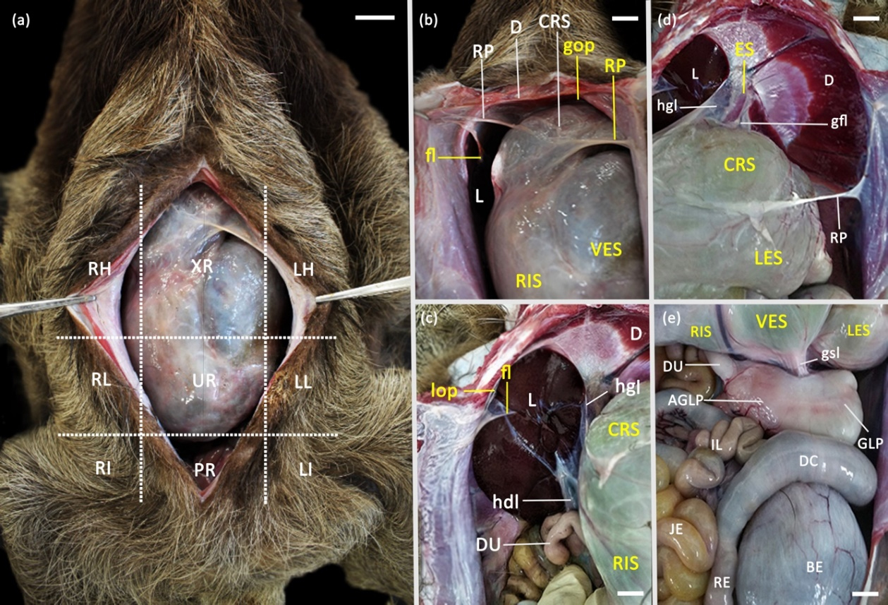
Figure 1
Photomacrography of the anatomical aspects of the stomach of Bradypus variegatus: (a) XR (xiphoid region), RH (right hypochondriacal region), LH (left hypochondriacal region), RL (right lateral abdominal region), LL (left lateral abdominal region), UR (umbilical region); RI (right inguinal region); LI (left inguinal region) and PR (pubic region); (b) Ventral visualization of the cranial boundaries and peritoneal folds of stomach attachment: CRS (cranial sac), VES (ventral sac), RIS (right lateral sac), RP (peritoneal recess), D (diaphragm), L (liver), gop (greater omental pouch), fl (falciform ligament); (c) Ventrolateral visualization of craniolateral syntopy and peritoneal folds that fix the organ dorsally: CRS (cranial sac), RIS (right lateral sac), D (diaphragm), L (liver), DU (duodenum), lop (lesser omental pouch), fl (falciform ligament), hgl (hepatogastric ligament), hdl (hepatoduodenal ligament); (d) Ventrolateral visualization of craniolateral syntopy and peritoneal folds that fix the organ dorsally and ventrolaterally: CRS (cranial sac), LES (left lateral sac), D (diaphragm), L (liver), ES (esophagus), RP (peritoneal recess), hgl (hepatogastric ligament), gfl (gastrophrenic ligament); (e) Ventrocaudal visualization of the boundaries and caudal syntopy of the stomach: RIS (right lateral sac), VES (ventral sac), LES (left lateral sac) GLP (glandular prepylorus), AGLP (aglandular prepylorus), DU (duodenum), IL (ileum), JE (jejunum), DC (descending colon), RE (rectum), BE (bladder), gsl (gastrosplenic ligament). Scale bar: 2cm.
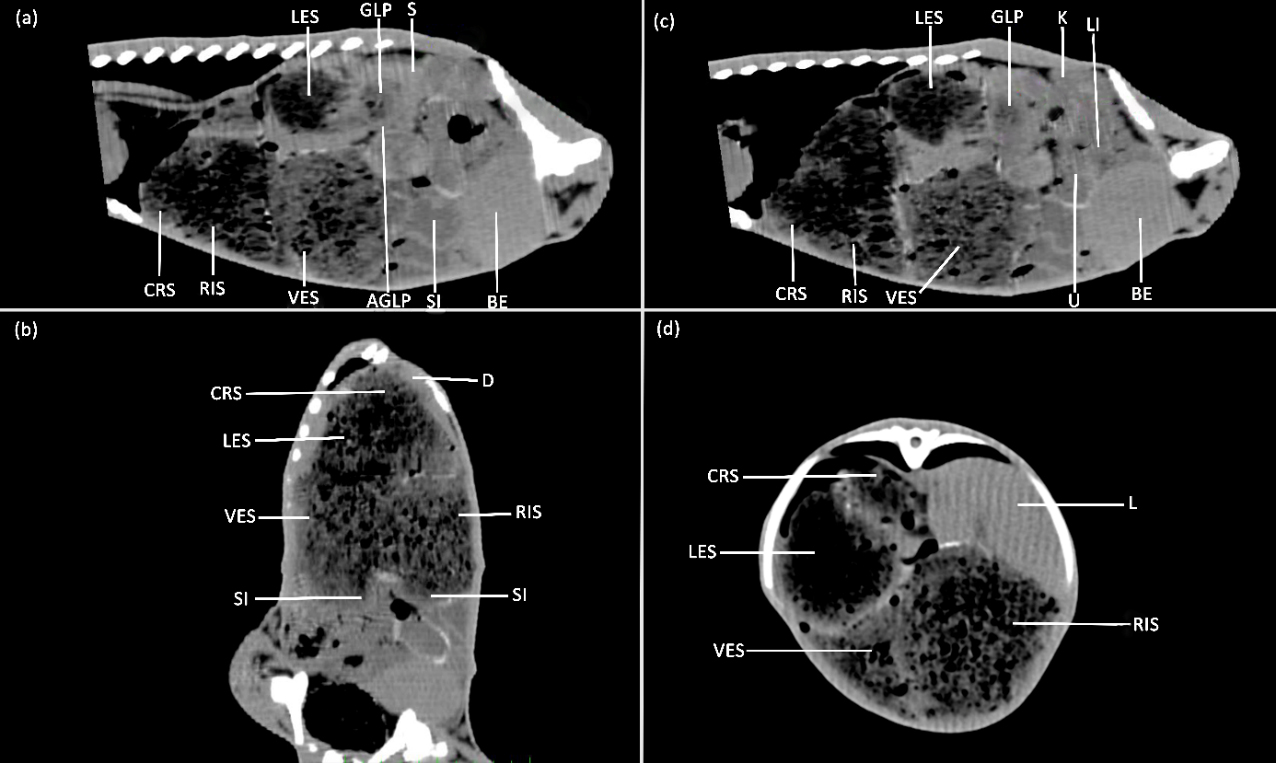
Figure 2
Tomography of the abdomen of Bradypus variegatus showing the topography and syntopy of the stomach: (a) CRS (cranial sac), LES (left lateral sac), VES (ventral sac), RIS (right lateral sac), GLP (glandular prepylorus), AGLP (aglandular prepylorus), S (spleen), SI (small intestine), BE (bladder); (b) Dorsal plane section of the abdomen: CRS (cranial sac), LES (left lateral sac), VES (ventral sac), RIS (right lateral sac), D (diaphragm), SI (small intestine); (c) Parasagittal section of the abdominal and pelvic cavity: CRS (cranial sac), LES (left lateral sac), VES (ventral sac), RIS (right lateral sac), GLP (glandular prepylorus), K (left kidney), LI (large intestine), U (uterus), BE (bladder); (d) Transverse section of the abdomen to the boundary of the 10th thoracic vertebra: CRS (cranial sac), LES (left lateral sac), VES (ventral sac), RIS (right lateral sac), L (liver).
Figure 3
Photomacrography of the external and internal morphology of the anatomical parts of the stomach of sloth Bradypus variegatus: (a) Ventral visualization; (b) Dorsal visualization, CRS (cranial sac), CRS’ (dorsal cranial sac part), LES (left lateral sac), VES (ventral sac), COS (connecting sac), RIS (right lateral sac), DIV (diverticulum), GLP (glandular prepylorus), AGLP (aglandular prepylorus), ES (esophagus), crasu (cranial sulcus), losu (longitudinal sulcus), cosu (coronary sulcus), acsu (accessory sulcus), casu (caudal sulcus); (c) Internal mucosa, CRS’ (dorsal cranial sac part), CRS’’(cranial sac ventral part), LES (left lateral sac), VES (ventral sac), COS (connecting sac), RIS (right lateral sac), DIV (diverticulum), ES (esophagus), crap (cranial pillar), caup (caudal pillar) lop (longitudinal pillar), cop (coronary pillar), acp (accessory pillar), car (cardia), GLP (glandular prepylorus), AGLP (aglandular prepylorus). (d) Internal mucosa, CRS’ (cranial sac dorsal part), LES (left lateral sac), VES (ventral sac), COS (connecting sac), RIS (right lateral sac), ES (esophagus), crap (cranial pillar), car (cardia), gasu (gastric sulcus); (e) Internal mucosa of the right lateral sac, RIS (right lateral sac), CRS’ (cranial sac dorsal part), VES (ventral sac), DIV (diverticulum), RISp (right lateral sac pouch), DIVp (diverticular folds); (f) Internal mucosa of the prepylorus, GLP (glandular prepylorus), AGLP (aglandular prepylorus), LES (left lateral sac), VS (ventricular sulcus), DUO (duodenum), Po (pyloric ostium). Scale bar: 3cm.
Figure 4
Photomicrography of the aglandular cardic stomach of Bradypus variegatus: (a) cranial sac: mucosa (M) with flat stratified keratinized epithelium (K) resting on a thin lamina proper of loose connective tissue, with absence of mucosal muscle, followed by a thin submucosa (SB) of connective tissue and thick layer of smooth muscle (MS) and thin serous layer (S); (b) left lateral sac: mucosa (M) with stratified epithelium with thick band of keratin, absence of mucosal muscle, thin submucosa (SB) and thin layer of smooth muscle (MS); (c) ventral sac: mucosa (M) lined by keratinized epithelium (K) with low stratification, thin submucosa (SB), dense layer of muscle tissue (MS) arranged in longitudinal, transverse and circular direction; (d) connecting sac: mucosa (M) of flat stratified and keratinized epithelium with lamina proper, thin submucosa (SB), thick muscle layer (MS) arranged in transverse and longitudinal direction. Scale bar (a): 200µm; (b): 100µm; (c): 50µm; (d): 200µm.
Figure 5
Photomicrography of the glandular cardic stomach of Bradypus variegatus: (a) right lateral sac: mucosa (M) with simple columnar epithelium of glandular tissue and mucus-producing cells, with dense mucosal muscularis, submucosa (SB) of loose connective tissue, muscular layer (MS) in double organization; (b) visualize transition (black arrows) of keratinized (K) aglandular epithelium of the cranial sac and glandular columnar epithelium mucosa (M) of the right lateral sac, mucosal muscularis slender (Mm), submucosa (SB) of loose connective tissue; (c) diverticulum: mucosa (M) of simple columnar epithelium with villi (V) projecting toward the lamina proper, followed by a dense mucosal muscularis (Mm), thick submucosa (SB), and still muscularis organized in two layers; (d) visualize the mucosa (M) villi projecting toward the lamina proper and mucosal muscularis slender (Mm). Scale bar (a): 100µm; (b): 200µm; (c): 200µm; (d): 50µm.
Figure 6
Photomicrography of the pyloric region of the stomach of Bradypus variegatus: (a) pre-pyloric glandular: mucosa (M) of simple columnar epithelium with glandular tissue forming the gastric pits, mucosal muscle (Mm) thin, submucosa (SB) of loose connective tissue and muscle layer (MS) dense; (b) visualize the oxyntic or parietal cells (black arrows) present in the upper half of the gastric glands and neck and zymogenic or principal cells (yellow arrows) contained in the lower half of the glands; (c) aglandular prepylorus: mucosa of keratinized stratified epithelium (K), submucosa (SB) of dense connective tissue and muscle (MS) with longitudinal and transverse arrangement; (d) visualize keratinized stratified epithelium (K) with a dense and thick band with lamina proper of loose connective tissue (black arrow), submucosa (SB) of loose connective tissue and muscular layer of smooth muscle (MS). Scale bar (a): 200µm; (b): 50µm; (c): 200µm; (d): 50µm.

 Contributions to study the anatomy of stomach of Bradypus variegatus (Mammalia: Folivora)
 Thumbnail
 Thumbnail
 Thumbnail
 Thumbnail
 Thumbnail
 Thumbnail