Contributions to study the anatomy of stomach of Bradypus variegatus (Mammalia: Folivora)
Abstract
Accurate anatomical and physiological knowledge of the stomach of the three-toed sloth is essential for effective clinical, surgical and dietary management, particularly in rehabilitation settings. This study analyzed the holotopy, topography, syntopy, and macro- and microscopic morphology of the stomach in thirteen B. variegatus specimens. Macroscopically, the stomach was located in the cranial, middle and abdominal regions and extended over the xiphoid, hypochondriac, lateral and umbilical regions. It had syntopic relationships with the diaphragm, liver, intestines, uterus (in females), vertebrae, ribs, spleen, pancreas, kidneys and abdominal musculature. The stomach showed distinct structural divisions, including saccular, diverticular and tubular segments, leading to the classification of seven anatomical regions: cranial sac, left and right lateral sacs, ventral sac, diverticulum, glandular pre-pylorus and non-glandular pre-pylorus. Histologically, the cranial sacs were non-glandular and keratinized, whereas the right lateral sac and diverticulum were glandular, the latter possibly involved in absorption. The cranial pre-pyloric region contained oxyntic and zymogenic glands, whereas the caudal pre-pyloric region lacked glands but had a keratinized mucosa. Unique anatomical features included peritoneal recesses, an omental pouch.
Key words
Xenarthra; morphology; foregut; sloth; Bradypodidae
INTRODUCTION
Sloths of the genus Bradypus Linnaeus, 1758 (Xenarthra) have an extensive distribution throughout the South American continent (Hayssen 2010), with emphasis on the species Bradypus variegatus Schinz, 1825, which occurs from Honduras in Central America, northern Colombia to the west and south of Venezuela, south in Ecuador, east of Peru and Bolivia, as well as the Brazilian Amazon and Atlantic Forest (Ruiz-García et al. 2020). Their main morphological characteristics include three digits on their forefeet, a brownish-yellow fur color, and a dark band around the eyes (Wetzel 1982). Regarding the aspects of the digestive system, they have eighteen molariform teeth with cusps (Albuquerque et al. 2016), a narrow esophagus located exclusively in the left antimer of the neck (Mesquita et al. 2019), intestines considered short for a herbivore (Rezende et al. 2011).
Regarding the anatomy of the stomach, classic studies have described the stomach of the sloth Bradypus spp. as a primitive organ (Britton 1941, Wislocki 1928) and over time different descriptions and classifications can be found for the stomach of bradypodids regarding the number of compartments and anatomical regions, with no agreement between the studies carried out to date (Bauchop & Martucci 1968, Rezende et al. 2011). More recently, based on histological data, it was suggested that this organ could have seven gastric compartments in B. variegatus living in the Brazilian Amazon (Mesquita et al. 2021).
Studies have demonstrated that the stomach is the main digestive organ in these animals, where fermentation occurs by symbiotic microorganisms, such as archaea, on the ingested food fractions (Dill-Mcfarland et al. 2016). On the other hand, the stomach is also reported to be the organ where most digestive disorders occur in ex situ management conditions, such as bloat and dysbiosis (Riaño et al. 2016), requiring knowledge of its anatomy for diagnosis and treatment, and in some cases, clinical-surgical interventions. However, detailed information about the anatomy of this organ is scarce (Dünner & Pastor 2017). Therefore, considering that the stomach is an essential organ in the digestion of sloths, the objective of this study was to provide a detailed description of the anatomy of the stomach of B. variegatus in order to provide information that can assist in semiological evaluations, diagnoses, and clinical-surgical interventions in this species.
MATERIALS AND METHODS
Animals
This study utilized thirteen adult cadavers of B. variegatus sloths, including seven males (5.5 kg) and six females (5.9 kg), donated to the anatomical collection of the Anatomy Department of the Federal Rural University of Pernambuco. Additionally, one live female sloth kept under semi-captive conditions at the Dois Irmãos Zoo in Recife, Brazil, was used for computed tomography analysis. All procedures were authorized by the Ethics Committee for Animal Experimentation of the Federal Rural University of Pernambuco, Recife, Brazil, under license number 033/2019, register Sisbio 46665-9 and Sisgen A0AFB89.
Macroscopic Description
The cadavers were dissected with usual instruments through a median sagittal incision of the skin, following the xiphoid process of the sternum bone up to the pubic region. The skin, ventral musculature and peritoneum were then retracted to expose the abdominal cavity and expose the stomach to allow description of the relationship of this organ to the body (holotopy), the position of the organ in the abdominal cavity (topography) and the relationship of the organ to its immediate neighbors (syntopy). To describe the morphology, the organ was clamped at the cardic and pyloric portions and removed from the abdominal cavity. It was then washed with running water and emptied of the gastric contents for macroscopic evaluation of the internal structures.
Microscopic Description
For the microscopic analysis, different anatomical parts of the stomach were sampled. They were washed with physiological solution and placed in containers for fixation in buffered formaldehyde for a period of 24 hours. Afterward, they were immersed in 70% alcohol for an additional 72 hours and dehydrated in increasing concentrations of ethanol. They were then embedded in paraffin (Leica Paraplast Plus) that was melted at 60°C. Once the blocks were formed, they were subjected to microtomy (Leica RM2125RT Microtome) to obtain 3μm histological sections, which were mounted on histological slides and stained using the hematoxylin-eosin (HE) method. The sections were examined under an optical microscope using the LASEZ 4.0 program (Leica).
Computed Tomography
A computed tomography (CT) scan was performed on a healthy, non-pregnant female sloth after a 12-hour fasting period for food and a 4-hour fasting period for water. The procedure was carried out using a single-channel GE CT scanner. The sloth was administered a sedative, Dexmedetomidine α-2, at a dosage of 10µg. The animal was positioned in a ventral decubitus position on the CT table, with the thoracic limbs displaced cranially and the pelvic limbs displaced caudally. The region evaluated extended from the chest to the coccygeal vertebrae to ensure that the entire abdominal and pelvic cavity was included in the examination and to facilitate visualization of the stomach.
The nomenclature adopted followed the standards of Nomina Anatomica Veterinaria (International Committee on Veterinary Gross Anatomical Nomenclature 2017) and Nomina Histologica Veterinaria (International Committee on Veterinary Gross Histological Nomenclature 2017).
RESULTS
The stomach of the sloth B. variegatus has three morphologically distinct segments: the cardic stomach, which includes the cranial, left lateral, ventral, and right lateral sacs; the gastric diverticulum; and the prepyloric stomach, which consists of the glandular prepylorus and the aglandular prepylorus, for a total of seven anatomical parts.
Holotopy
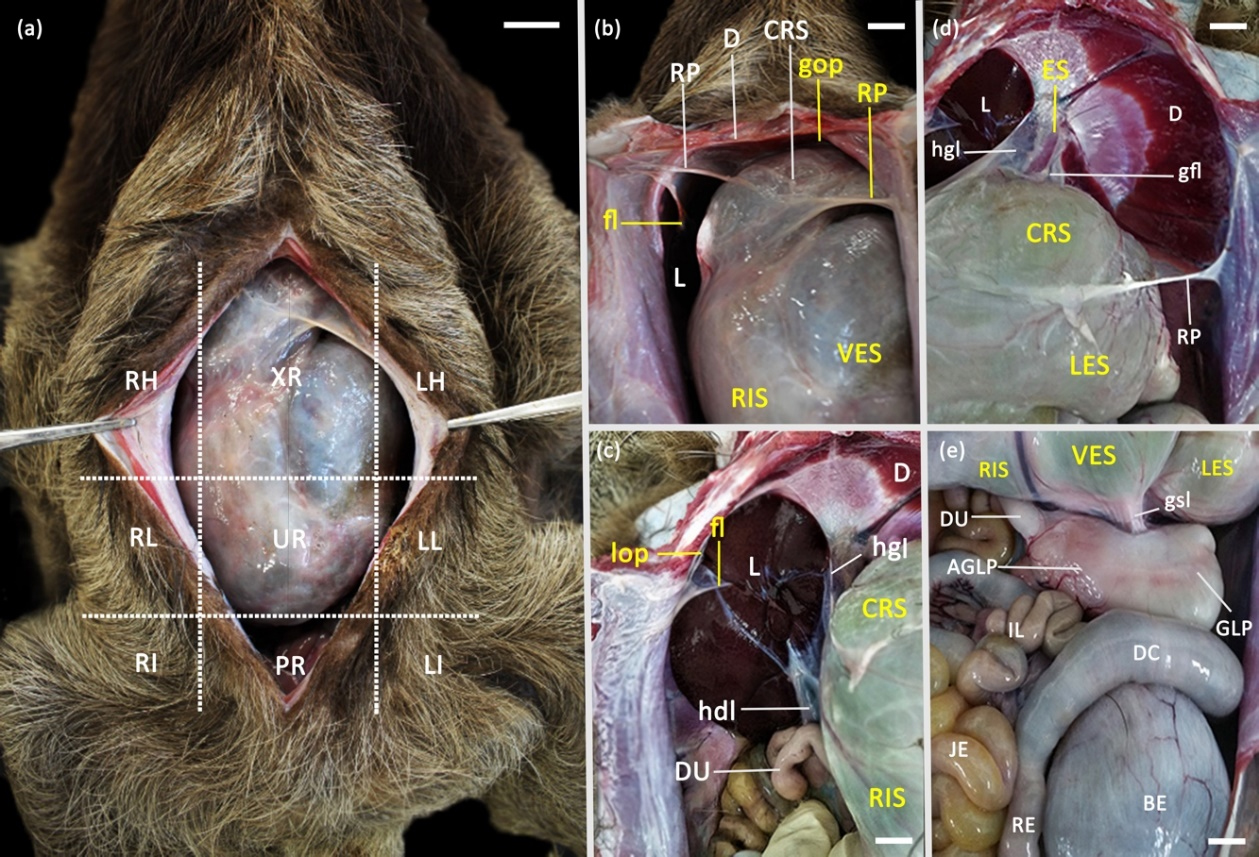
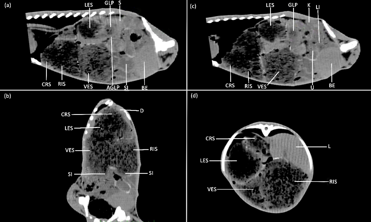
Dissection showed a large stomach in the abdomen of males and females, predominantly located in the cranial region and a small portion in the middle region of the abdominal cavity, extending completely from the left antimere to the right in all specimens evaluated (Figure 1a and 2b). It was bordered cranially by the diaphragm and liver in the intrathoracic space, caudally by the small and large intestines, and also by the uterus in the females (Figure 1b, c and 2b, c). Laterally it is bordered by the costal arches on its medial side and intercostal muscles from the eighth to the fifteenth rib on the left antimere and between the tenth and the fifteenth rib on the right antimere (Figure 2a, b). Dorsally it is bounded between the eighth thoracic and second lumbar vertebrae, spleen and kidneys and ventrally it is bounded by the internal abdominal oblique and rectus abdominis muscles (Figure 1a, 2a, d).

Photomacrography of the anatomical aspects of the stomach of Bradypus variegatus: (a) XR (xiphoid region), RH (right hypochondriacal region), LH (left hypochondriacal region), RL (right lateral abdominal region), LL (left lateral abdominal region), UR (umbilical region); RI (right inguinal region); LI (left inguinal region) and PR (pubic region); (b) Ventral visualization of the cranial boundaries and peritoneal folds of stomach attachment: CRS (cranial sac), VES (ventral sac), RIS (right lateral sac), RP (peritoneal recess), D (diaphragm), L (liver), gop (greater omental pouch), fl (falciform ligament); (c) Ventrolateral visualization of craniolateral syntopy and peritoneal folds that fix the organ dorsally: CRS (cranial sac), RIS (right lateral sac), D (diaphragm), L (liver), DU (duodenum), lop (lesser omental pouch), fl (falciform ligament), hgl (hepatogastric ligament), hdl (hepatoduodenal ligament); (d) Ventrolateral visualization of craniolateral syntopy and peritoneal folds that fix the organ dorsally and ventrolaterally: CRS (cranial sac), LES (left lateral sac), D (diaphragm), L (liver), ES (esophagus), RP (peritoneal recess), hgl (hepatogastric ligament), gfl (gastrophrenic ligament); (e) Ventrocaudal visualization of the boundaries and caudal syntopy of the stomach: RIS (right lateral sac), VES (ventral sac), LES (left lateral sac) GLP (glandular prepylorus), AGLP (aglandular prepylorus), DU (duodenum), IL (ileum), JE (jejunum), DC (descending colon), RE (rectum), BE (bladder), gsl (gastrosplenic ligament). Scale bar: 2cm.

Tomography of the abdomen of Bradypus variegatus showing the topography and syntopy of the stomach: (a) CRS (cranial sac), LES (left lateral sac), VES (ventral sac), RIS (right lateral sac), GLP (glandular prepylorus), AGLP (aglandular prepylorus), S (spleen), SI (small intestine), BE (bladder); (b) Dorsal plane section of the abdomen: CRS (cranial sac), LES (left lateral sac), VES (ventral sac), RIS (right lateral sac), D (diaphragm), SI (small intestine); (c) Parasagittal section of the abdominal and pelvic cavity: CRS (cranial sac), LES (left lateral sac), VES (ventral sac), RIS (right lateral sac), GLP (glandular prepylorus), K (left kidney), LI (large intestine), U (uterus), BE (bladder); (d) Transverse section of the abdomen to the boundary of the 10th thoracic vertebra: CRS (cranial sac), LES (left lateral sac), VES (ventral sac), RIS (right lateral sac), L (liver).
The anatomical position of the stomach is maintained in the abdominal cavity by peritoneal folds originating from the dorsal mesogastric such as the gastrophrenic and gastrosplenic ligament and also by peritoneal folds from the ventral mesogastric such as the hepatoduodenal and hepatogastric ligaments and a thin lamina that continues to the falciform ligament, which in turn attaches ventrolaterally to the abdominal wall (Figure 1b, c, d, e). Extensive peritoneal folds are also noted that attach this organ ventrolaterally (Figure 1b, d). A space without peritoneal connection was observed between the cranial margin of the cranial sac, left tendinous center of the diaphragm and the diaphragmatic muscle portion, forming an omental cavity; however, this region was collapsed upon dissection and could only be observed through incision. The organ was again attached more laterally by peritoneal folds that fused to the lateral wall of the abdomen (Figure 1b, d).
Topography
The cranial sac of the stomach was located in the xiphoid region, deviating to the left hypochondriac region, comprising the intercostal spaces between the seventh and tenth ribs (Figure 1a). The left lateral sac was located between the xiphoid and left hypochondriac regions, meeting at the limits between the tenth and thirteenth ribs, covered by a small layer of lateral abdominal muscles (Figure 1c). The ventral sac was positioned between the xiphoid and umbilical region, deviating to the left hypochondriacal region from the median plane (Figure 1c). The right lateral sac was located between the xiphoid, right hypochondriacal and right abdominal regions, between the eleventh and fifteenth rib spaces and caudally in the umbilical region (Figure 1b).
The diverticulum originates in the umbilical region, projecting transversely into the left abdominal region, with its conical apex ascending cranioventrally into the left hypochondriac region, resting on a groove between the left lateral sac and ventral sac and on the glandular and aglandular prepylorus. The glandular prepylorus was located in the left hypochondriacal region in the space between the tenth and twelfth ribs, and the aglandular prepylorus was located between the left hypochondriacal and left abdominal regions in a craniocaudal arrangement at the boundaries between the twelfth and fifteenth rib costal spaces, with its end portion projecting transversely to the duodenum in the median plane (Figure 1e).
Sintopy
The cranial sac of the stomach maintains syntopy cranially with esophagus in the cardic region, diaphragm and dorsally with the thoracic vertebrae (T7 to T10) (Figure 1b, c, d). The left lateral sac has syntopic relationship with intercostal muscles between tenth and thirteenth ribs laterally (Figure 1d, e). The ventral sac syntopically relates to abdominal muscles ventrally (Figure 1b, e). The right lateral sac has a relationship with the liver cranially, the intercostal muscles of the eleventh and fifteenth ribs and small intestine caudally (Figure 1b, c, e) and the diverticulum has a relationship caudally with the small and large intestines. The glandular prepylorus syntopses with the internal musculature of the tenth to twelfth ribs, descending colon and spleen via the gastrosplenic ligament while the aglandular prepylorus syntopses dorsally with pancreas, caudally with ileum and medially with duodenum (Figure 1e).
External macroscopic morphology
The stomach is a pluricavitary organ and externally shows large sacculiform dilations, having circular and concave borders in the presence of digesta, but at the caudal end of the right lateral sac it narrows into a diverticular sac forming a long conical appendix and the final third of the stomach shows as a fusiform segment divided into two parts by a medial constriction, which precedes the pylorus and duodenum, called in this study pre-pylorus glandular and aglandular (Figure 3a, b). The main sacculations are delimited internally by pillars, some of which can be seen in the serosa of the stomach as grooves, although they are not very pronounced on the external surface. A cranial pillar delimits the cranial sac and the left lateral sac ventrally and a caudal pillar demarcates them caudo-dorsally. A longitudinal pillar divides the cranial sac and the right lateral sac, which is not directly separated from the diverticulum; a coronary groove delimits and surrounds the ventral sac, separating it from the right lateral sac and diverticulum. An accessory pillar delimits the left lateral sac (Figure 3c). At the junction between the left lateral sac and the glandular prepylorus there are no obvious grooves, but between the latter and the aglandular prepylorus there is a medial constriction (Figure 3a, b).

Photomacrography of the external and internal morphology of the anatomical parts of the stomach of sloth Bradypus variegatus: (a) Ventral visualization; (b) Dorsal visualization, CRS (cranial sac), CRS’ (dorsal cranial sac part), LES (left lateral sac), VES (ventral sac), COS (connecting sac), RIS (right lateral sac), DIV (diverticulum), GLP (glandular prepylorus), AGLP (aglandular prepylorus), ES (esophagus), crasu (cranial sulcus), losu (longitudinal sulcus), cosu (coronary sulcus), acsu (accessory sulcus), casu (caudal sulcus); (c) Internal mucosa, CRS’ (dorsal cranial sac part), CRS’’(cranial sac ventral part), LES (left lateral sac), VES (ventral sac), COS (connecting sac), RIS (right lateral sac), DIV (diverticulum), ES (esophagus), crap (cranial pillar), caup (caudal pillar) lop (longitudinal pillar), cop (coronary pillar), acp (accessory pillar), car (cardia), GLP (glandular prepylorus), AGLP (aglandular prepylorus). (d) Internal mucosa, CRS’ (cranial sac dorsal part), LES (left lateral sac), VES (ventral sac), COS (connecting sac), RIS (right lateral sac), ES (esophagus), crap (cranial pillar), car (cardia), gasu (gastric sulcus); (e) Internal mucosa of the right lateral sac, RIS (right lateral sac), CRS’ (cranial sac dorsal part), VES (ventral sac), DIV (diverticulum), RISp (right lateral sac pouch), DIVp (diverticular folds); (f) Internal mucosa of the prepylorus, GLP (glandular prepylorus), AGLP (aglandular prepylorus), LES (left lateral sac), VS (ventricular sulcus), DUO (duodenum), Po (pyloric ostium). Scale bar: 3cm.
Internal Macroscopic Morphology
On the inner face inflections of pillars that delimit the major gastric sacculations and make the organ compartmentalized are visualized. These inflections are visible on the external face as grooves already described that correspond to the position of the pillars. Internally, the cranial, left lateral, and ventral sacs macroscopically exhibited darker, slightly wrinkled, and folded mucosa (Figure 3c, d). The right lateral sac and diverticulum showed smoother and clearer surface and the right lateral sac exposed even in the transition with the diverticulum small folds facing the lumen that originate pouches, as well as the diverticulum exhibited laminar folds that formed small compartments (Figure 3c, e). The glandular prepylorus revealed a predominantly smooth and thin mucosa and its cranial portion with the left lateral sac has a rough and pleated ventricular groove, while the aglandular prepylorus has a wrinkled, rough and darkened mucosa (Figure 3f).
Histology
The cranial sac of the stomach showed a mucosa consisting of a flat stratified keratinized epithelium over a thin lamina propria of loose connective tissue without the presence of mucosal muscle, continued by the submucosa formed of a thin layer of dense connective tissue and deeper by a thick layer of smooth muscle and followed by a serous layer of connective tissue (Figure 4a). The left lateral sac of the stomach revealed the same histological constitution as the cranial sac, also lacking the mucosal muscle, but possessing a thick band of keratin (Figure 4b). The ventral sac also exhibited a mucosa lined by keratinized epithelium with low stratification, thin submucosa, followed by a dense layer of smooth muscle tissue arranged in a longitudinal, transverse and circular direction, followed by a dense connective tissue serosa (Figure 4c). The connecting region between the sacs showed flat, keratinized stratified epithelium with lamina propria, thin submucosa followed by a thick layer of muscle arranged in transverse and longitudinal direction and loose connective tissue serosa (Figure 4d).

Photomicrography of the aglandular cardic stomach of Bradypus variegatus: (a) cranial sac: mucosa (M) with flat stratified keratinized epithelium (K) resting on a thin lamina proper of loose connective tissue, with absence of mucosal muscle, followed by a thin submucosa (SB) of connective tissue and thick layer of smooth muscle (MS) and thin serous layer (S); (b) left lateral sac: mucosa (M) with stratified epithelium with thick band of keratin, absence of mucosal muscle, thin submucosa (SB) and thin layer of smooth muscle (MS); (c) ventral sac: mucosa (M) lined by keratinized epithelium (K) with low stratification, thin submucosa (SB), dense layer of muscle tissue (MS) arranged in longitudinal, transverse and circular direction; (d) connecting sac: mucosa (M) of flat stratified and keratinized epithelium with lamina proper, thin submucosa (SB), thick muscle layer (MS) arranged in transverse and longitudinal direction. Scale bar (a): 200µm; (b): 100µm; (c): 50µm; (d): 200µm.
The right lateral sac of the stomach revealed glandular tissue mucosa with mucus producing cells in simple columnar epithelium, with a thin muscularis mucosa, followed by a loose connective tissue submucosa, muscularis layer organized in two bands, continuing with a loose connective tissue serosa (Figure 5a, b). The diverticulum of the stomach has in its mucosa villi lined by simple columnar epithelium that project toward its lamina propria, followed by a dense mucosal muscularis, dense and thick submucosa, and further muscularis organized in two layers and dense serosa more externally (Figure 5c, d). In the glandular prepylorus a mucosa with gastric foveolar invaginations consisting of oxyntic and zymogenic cells was observed. The mucosa is arranged by a thin lamina propria of loose connective tissue and adjacent to this the mucosal muscle layer can be observed, followed by thin submucosa and dense muscle layer (Figure 6a, b). The aglandular prepylorus showed keratinized stratified epithelium and loose connective tissue lamina propria, thick dense connective tissue submucosa, and longitudinally and transversely organized muscle layer (Figure 6c, d).

Photomicrography of the glandular cardic stomach of Bradypus variegatus: (a) right lateral sac: mucosa (M) with simple columnar epithelium of glandular tissue and mucus-producing cells, with dense mucosal muscularis, submucosa (SB) of loose connective tissue, muscular layer (MS) in double organization; (b) visualize transition (black arrows) of keratinized (K) aglandular epithelium of the cranial sac and glandular columnar epithelium mucosa (M) of the right lateral sac, mucosal muscularis slender (Mm), submucosa (SB) of loose connective tissue; (c) diverticulum: mucosa (M) of simple columnar epithelium with villi (V) projecting toward the lamina proper, followed by a dense mucosal muscularis (Mm), thick submucosa (SB), and still muscularis organized in two layers; (d) visualize the mucosa (M) villi projecting toward the lamina proper and mucosal muscularis slender (Mm). Scale bar (a): 100µm; (b): 200µm; (c): 200µm; (d): 50µm.

Photomicrography of the pyloric region of the stomach of Bradypus variegatus: (a) pre-pyloric glandular: mucosa (M) of simple columnar epithelium with glandular tissue forming the gastric pits, mucosal muscle (Mm) thin, submucosa (SB) of loose connective tissue and muscle layer (MS) dense; (b) visualize the oxyntic or parietal cells (black arrows) present in the upper half of the gastric glands and neck and zymogenic or principal cells (yellow arrows) contained in the lower half of the glands; (c) aglandular prepylorus: mucosa of keratinized stratified epithelium (K), submucosa (SB) of dense connective tissue and muscle (MS) with longitudinal and transverse arrangement; (d) visualize keratinized stratified epithelium (K) with a dense and thick band with lamina proper of loose connective tissue (black arrow), submucosa (SB) of loose connective tissue and muscular layer of smooth muscle (MS). Scale bar (a): 200µm; (b): 50µm; (c): 200µm; (d): 50µm.
DISCUSSION
Macroscopic aspects
Among the main findings of this study we can highlight the identification of some previously undocumented anatomical structures for the stomach of B. variegatus, such as the number of pillars that divide the saccular segmentations, called in this study the cranial sac, left lateral sac, ventral sac, right lateral sac, which added to the gastric diverticulum, glandular pre-pylorus and aglandular pre-pylorus give the organ a total of seven anatomical parts. These results corroborate the descriptions, at least in terms of the number of anatomical regions, reported in a previous study of B. variegatus by Mesquita et al. (2021) and for B. torquatus (Rezende et al. 2011).
The topographic position of this organ in the abdomen resembled the large stomachs found in Ruminantia (Langer 1974) and Tylopoda (Vater et al. 2021). However, by extending integrally from the right to the left antimer in the cranial and middle regions of the abdomen, the stomach of B. variegatus fills a space, in proportion, even larger compared to the aforementioned ruminants, where the stomachs occupy half of the left antimer and only part of the right antimer in the same topographic regions of the abdomen (Hofmann 1989, Vater et al. 2021).
The peritoneal recesses that attach this organ ventrolaterally to the abdominal wall seem to be a characteristic feature of these animals, since in other mammals most of the attachment of this organ is by a well visible greater omentum that originates dorsally on the roof of the abdominal cavity and projects as a lamina over the stomach (König & Liebich 2021). We also noted the existence of a cavity between the cranial margin of the cranial sac, left tendinous center of the diaphragm and diaphragmatic muscle portion and proposed that this space actually represents the omental pouch, disagreeing with previous studies that reported absence of omentum major for sloths of this genus (Rezende et al. 2011, Mesquita et al. 2021).
The distended saccules of the stomach were similar to saculiform dilations described for colobids (Bauchop & Martucci 1968, Stevens & Hume 1995) and ruminants (Hofmann 1989, Machado et al. 2015), however it differs from hippos and camelids, whose stomach saccules are elongated and sigmoidal (Langer 1984, Stevens & Hume 1995). Such sacculations are bordered by transverse and longitudinal grooves in a similar manner to the proventricles of ruminants (Machado et al. 2015, König & Liebich 2021). The conical diverticular segment has been shown to be like the tubiform appendage of the saciform stomach of red kangaroos (Shoeib et al. 2015) and in the compartmentalized stomach of manatees and dugongs (Stevens & Hume 1995). The fusiform segment preceding the pylorus and duodenum, similar to that observed in domestic and wild animals (Stevens & Hume 1995, König & Liebich 2021), but in B. variegatus there is a constriction dividing this region into two parts, as observed in the two-toed sloth Choloepus sp. (Wislocki 1928).
The pillar inflections delimiting the largest gastric sacculations can be compared to those described for wild ruminants (Machado et al. 2015). However, the lumen-facing folds that give rise to small pouches in the right lateral sac and diverticulum had not yet been reported as a characteristic feature of B. variegatus, as such a morphological attribute is uncommon to other pluricavitaries (Machado et al. 2015). Another striking feature is the absence of macropapillae in all gastric sacculations which differs from that observed in domestic and wild ruminants, which have in their gastric chambers structures such as ridges, laminar folds and very prominent papillae.
Microscopic aspects
The flat keratinized stratified epithelium found in the cranial, left lateral and ventral sacculae resembled the gastric chambers of ruminants, aglandular cardic region of horses and pigs (König & Liebich 2021) and kangaroos (Shoeib et al. 2015). In known ruminants, this type of tissue found in saccules performs protective functions against mechanical injury from fibrous foods and are the chambers of bacterial fermentation for digestion of structural carbohydrates such as cellulose, which constitutes a large part of their diets (Clauss et al. 2009), a characteristic that can be sustained for B. variegatus, since they feed strictly on leaves rich in cellulose (Cliffe et al. 2015). In addition, studies have demonstrated high fermentative activity in these anatomical regions in Bradypus sloths (Foley et al. 1995).
In contrast, the right lateral sac showed a mucosa of glandular tissue with mucus-producing cells characteristic of a cardic glandular stomach (Langer 1984), for this reason, we propose to call this structure the right cardic sac, in contrast to the one used for B. variegatus by Mesquita et al. (2021), called the fundic sac, because no oxyntic (parietal) or zymogenic (main) cells were visualized in this gastric chamber, characteristic of a fundic stomach (Langer 1984, König & Liebich 2021). In the cardic region of the mammalian stomach, mucus-producing cells secrete mucinogen and bicarbonate ions to protect the gastric lining from damage by an acidic pH and also to keep it more alkaline for digestive functions (König & Liebich 2021). We propose that the latter seems to be the most plausible function of this anatomical region in B. variegatus, since the pH of the stomach chambers in Bradypus sp. has a value close to 6, benefiting cellulose and hemicellulose fermenting bacteria (Foley et al. 1995). In addition, sloths do not have a large secretion of saliva (Quintarelli & Dellovo 1969) which commonly contributes to maintaining the pH alkalinity in the proventricles in other pregastric fermenters (Clauss & Hummel 2017).
These cells are also recognized for secreting lysozymes in the mammalian stomach (Jollès et al. 1989), and in the case of gastric fermenters, modified for the digestive function of the cell body of bacteria in the acid stomach (Dobson et al. 1984). However, based on the large number of glands in this region of the cardic stomach, we agree with the propositions of Pacheco et al. (2007) who suggested that the lysozymes of this gastric chamber in B. variegatus play more of a defensive role against foreign bacteria than a digestive role. The authors stated that this defense role maintains the balance in the symbiotic bacterial community, since their isoforms showed specific enzymatic activity three times greater in the aglandular sacculations than in the acid glandular pre-pylorus, especially at pH between 5.5 and 7.7.
The large villus cells in the mucosa of the diverticulum in B. variegatus may be associated with the absorption of volatile fermentation acids from the more cranial chambers. The structures of the diverticulum and its functions are not clear for sloths, where the most accepted role is related to a greater capacity to retain the digesta (Nagy & Montgomery 1980), but physiological studies in other species show that the diverticulum always contains fermentation gases and occasionally ingesta (Bauchop & Martucci 1968), supporting our propositions. In addition, internally this appendix exhibits some folds that resemble the laminar projections found in what constitutes the omasum of ruminants specialized in the absorption of volatile acids (Peters et al. 1990).
The presence of oxyntic and zymogenic cells in the first fusiform segment of the stomach suggests that this region can be considered the proper gastric stomach in B. variegatus, a constitution similar to the fundic stomach of mammals in general (Eurell & Frappier 2006). In contrast to this, the second fusiform stomach segment revealed an aglandular mucosa similar to the constitution of the cranial sacculae described above. Despite the similarity to a keratinized region of the pylorus of pangolins (Xu et al. 2020), we did not find an analogous histological arrangement in any other herbivorous mammal that could be compared to sloths.
Even among the other xenarthras such as anteaters, where a thickening of the muscle layer occurs, the pylorus is not keratinized and there is formation of the long gastric pits and mucus-secreting cells (Pinheiro et al. 2014). Or even in the armadillo that presents a muscular tubiform narrowing of the pylorus but mucosa consisting of mucus-producing cells, oxyntic and zymogenic, but without keratinization (Cavalcante et al. 2021), which makes this anatomical region in the sloth B. variegatus unique to these animals and must be related to their feeding habit that is mostly based on leaves.
CONCLUSIONS
This study revealed that the stomach of the sloth B. variegatus is segmented and can be divided into seven anatomical parts, occupying all the topographic regions of the cranial and middle portion of the abdomen, pointing to the significance of the digestive processes in these animals. Our findings showed unique macroscopic structures such as lateral peritoneal recesses for organ attachment, omental pouch, diverticular folds, and microscopically large villi in the diverticulum that may be associated with the absorption of fermentation products.
Acknowledgements
The authors would like to express their gratitude to the Anatomy Department of the Federal Rural University of Pernambuco for generously providing the specimens from their anatomical collection, which were dissected and evaluated in this study.
References
-
ALBUQUERQUE PV, SANTOS FC, GALVÃO APO, JÚNIOR FCAA, ROSAS EP & D’EMERY MB. 2016. Morphological analysis of teeth in Bradypus variegatus Schinz, 1825 (Mammalia, Bradypodidae). Biot 29: 35-40. https://doi.org/10.5007/2175-7925.2016v29n3p35.
» https://doi.org/10.5007/2175-7925.2016v29n3p35 -
BAUCHOP T & MARTUCCI RW. 1968. Ruminant-like digestion of the langur monkey. Scie 167: 698-700. https://doi.org/10.1126/science.161.3842.698.
» https://doi.org/10.1126/science.161.3842.698 - BRITTON SW. 1941. Form and function in the sloth. Quart Rev of Biol 16: 196-207. Available at: https://www.jstor.org/stable/2808832.
-
CAVALCANTE MMAS, FONSECA CMB, SILVA ABS, MENDONÇA TGS, RIZZO MS, LEITE CMC, PALOMBIT K & CONDE JÚNIOR AM. 2021. Structural cells identification in armadillo stomach: an opportunity to know about its digestion. Inter J Dev Res 11: 432-439. https://doi.org/10.37118/ijdr.20864.01.2021.
» https://doi.org/10.37118/ijdr.20864.01.2021 -
CLAUSS M, FRITZ J, BAYER D, NYGREN K, HAMMER S, HATT JM, SÜDEKUM KH & HUMMEL J. 2009. Physical characteristics of rumen contents in four large ruminants of different feeding type, the addax (Addax nasomaculatus), bison (Bison bison), red deer (Cervus elaphus) and moose (Alces alces). Comp Bio and Phy A 152: 398-406. https://doi.org/10.1016/j.cbpa.2008.11.009.
» https://doi.org/10.1016/j.cbpa.2008.11.009 -
CLAUSS M & HUMMEL J. 2017. Physiological adaptations of ruminants and their potential relevance for production systems. Rev Bras Zootec 46: 606-613. https://doi.org/10.1590/S1806-92902017000700008.
» https://doi.org/10.1590/S1806-92902017000700008 -
CLIFFE RN, HAUPT RJ, AVEY-ARROYO JÁ & WILSON RP. 2015. Sloths like it hot: ambient temperature modulates food intake in the brown- throated sloth (Bradypus variegatus). Peer 2: e875. http://dx.doi.org/10.7717/peerj.875.
» https://doi.org/10.7717/peerj.875 -
DILL-MCFARLAND KA, WEIMER PJ, PAULI JN, PEERY MZ & SUEN G. 2016. Diet specialization selects for an unusual and simplified gut microbiota in two- and three-toed sloths. Envir Microb 18: 1391-1402. https://doi.org/10.1111/1462-2920.13022.
» https://doi.org/10.1111/1462-2920.13022 -
DOBSON DE, PRAGER EM & WILSON AC. 1984. Stomach lysozymes of ruminants. Jour Biol Chem 259: 11607-11616. https://doi.org/10.1016/S0021-9258(18)90907-5.
» https://doi.org/10.1016/S0021-9258(18)90907-5 - DÜNNER C & PASTOR G. 2017. Manual de manejo, medicina y rehabilitación de perezosos. Fundación Huálamo, 1ª ed., Fundación Huálamo, Chile, 78 p.
- EURELL J & FRAPPIER BL. 2006. The digestive system. In: Dellmann’s Textbook of Veterinary Histology, Iowa, 6ª ed., Wiley-Blackwell, 176 p.
-
FOLEY WJ, ENGELHARDT WV & CHARLES-DOMINIQUE P. 1995. The passage of digesta, particle size, and in vitro fermentation rate in the three-toed sloth Bradypus tridactylus (Edentata: Bradypodidae). J Zool 236: 681-696. https://doi.org/10.1111/j.1469-7998.1995.tb02739.x.
» https://doi.org/10.1111/j.1469-7998.1995.tb02739.x -
HAYSSEN V. 2010. Bradypus variegatus (Pilosa: Bradypodidae). Mamm Spec 42: 19-32. https://doi.org/10.1644/850.1.
» https://doi.org/10.1644/850.1 - HOFMANN RR. 1989. Evolutionary steps of ecophysiological adaptation and diversification of ruminants: a comparative view of their digestive system. Oecol 78: 443-457.
-
INTERNATIONAL COMMITTEE ON VETERINARY GROSS ANATOMICAL NOMENCLATURE. 2017. Nomina Anatomica Veterinaria. 6th ed., World Association of Veterinary Anatomists, 178 p. Available at: https://www.wava-amav.org/wava-documents.html
» https://www.wava-amav.org/wava-documents.html -
INTERNATIONAL COMMITTEE ON VETERINARY GROSS HISTOLOGICAL NOMENCLATURE. 2017. Nomina Histologica Veterinaria. World Association of Veterinary Anatomists, 78 p. Available at: https://www.wava-amav.org/wava-documents.html
» https://www.wava-amav.org/wava-documents.html -
JOLLÈS J, JOLLÈS P, BOWMAN BH, PRAGER EM & STEWART C. 1989. Episodic evolution in the stomach lysozymes of ruminants. J Molec Evol 28: 528-535. https://doi.org/10.1007/BF02602933.
» https://doi.org/10.1007/BF02602933 - KÖNIG HE & LIEBICH HG. 2021. Anatomia dos Animais Domésticos: Texto e Atlas Colorido, Artmed, 7ª ed., 856 p.
-
LANGER P. 1974. Stomach evolution in the Artiodactyla. Mamm 38: 295-314. https://doi.org/10.1515/mamm.1974.38.2.295.
» https://doi.org/10.1515/mamm.1974.38.2.295 -
LANGER P. 1984. Comparative anatomy of the stomach in mammalian herbivores. Quart Jour of Exp Phys 69: 615-625. https://doi.org/10.1113/expphysiol.1984.sp002848.
» https://doi.org/10.1113/expphysiol.1984.sp002848 -
MACHADO MRF, LEAL LM, SASAHARA THC, OLIVEIRA FS & DUARTE JMB. 2015. Morfologia do estômago do cervo-do-pantanal (Blastocerus dichotomus, Illiger 1815). Arq Bras Med Veter Zootec 67: 424-432. https://doi.org/10.1590/1678-7366.
» https://doi.org/10.1590/1678-7366 -
MESQUITA EYE, SOARES PC, MELLO LR, FREIRE ECB & LIMA AR. 2021. Sloths (Bradypus variegatus) as a polygastric mammal. Micro Res & Techn 84: 79-88. https://doi.org/10.1002/jemt.23568.
» https://doi.org/10.1002/jemt.23568 -
MESQUITA EYE, SOARES PC, MELLO LR, LIMA AR, GIESE EG & BRANCO E. 2019. Morfologia do esôfago de Bradypus variegatus (Schinz, 1825). Biote 32: 97-104. https://doi.org/10.5007/2175-7925.2019v32n3p97.
» https://doi.org/10.5007/2175-7925.2019v32n3p97 -
NAGY KA & MONTGOMERY GG. 1980. Field metabolic rate, water flux, and food intake in three-toed sloths (Bradypus variegatus). J Mamm 61: 465-472. https://doi.org/10.2307/1379840.
» https://doi.org/10.2307/1379840 -
PACHECO MA, CONCEPCIÓN JL, RANGEL JDR, RUIZ MC & MICHELANGELI F. 2007. Stomach lysozymes of the three-toed sloth (Bradypus variegatus), an arboreal folivore from the neotropics. Comp Bioch Phys 147: 808-819. https://doi.org/10.1016/j.cbpa.2006.07.010.
» https://doi.org/10.1016/j.cbpa.2006.07.010 -
PETERS JP, SHEN RYW & CHESTER ST. 1990. Propionic acid disappearance from the foregut and small intestine of the beef steer. J Anim Scie 68: 3905-3913. https://doi.org/10.2527/1990.68113905x.
» https://doi.org/10.2527/1990.68113905x -
PINHEIRO ACO, LIMA AR, CARVALHO AF, PEREIRA LC & BRANCO E. 2014. Macroscopic and microscopic morphological aspects of lesser anteater stomach (Tamandua tetradactyla). Arq Bras Medic Vet Zootec 66: 1089-1096. https://doi.org/10.1590/1678-7140.
» https://doi.org/10.1590/1678-7140 -
QUINTARELLI G & DELLOVO MC. 1969. Studies on the exocrine secretions. Histoch 19: 199-223. https://doi.org/10.1007/BF00305284.
» https://doi.org/10.1007/BF00305284 - REZENDE LC, MONTEIRO JM, CARVALHO P, FERREIRA JR & MIGLINO MA. 2011. Morphology and vascularization of the gastric compartments in three-toed sloth (Bradypus torquatus Illiger, 1811). Inter J Morph 29: 1282-1290. Available at: https://www.scielo.cl/pdf/ijmorphol/v29n4/art35.pdf.
-
RIAÑO DMD, VILLAMIZAR NP & RICO CIB. 2016. Casuística de perezosos (Bradypus variegatus y Choloepus hoffmanni) recibidos en la Unidad de Rescate y Rehabilitación de Animales Silvestres de la Universidad Nacional de Colombia entre 2005 y 2010. Edent 17: 34-40. https://doi.org/10.2305/IUCN.CH.2016.EDENTATA-17-1.6.en.
» https://doi.org/10.2305/IUCN.CH.2016.EDENTATA-17-1.6.en - RUIZ-GARCÍA M, CHACÓN D, PLEASE T & SHOSTELL JM. 2020. Molecular phylogenetics of Bradypus (Three-Toed Sloth, Pilosa: Bradypodidae, Mammalia) and phylogeography of Bradypus variegatus (Brown-Throated Three-Toed Sloth) with Mitochondrial Gene Sequences. J Mamm Evol 27: 461-482. Available at: https://link.springer.com/article/10.1007/s10914-019-09465-w.
-
SHOEIB MB, HASSANIN A & ELNASHARTY M. 2015. Morphological and morphometric characteristics of gastric mucosa in western grey kangaroo (Macropus fuliginosus). J Adv Vet an Anim Res 2: 40-48. https://doi.org/10.5455/javar.2015.b43.
» https://doi.org/10.5455/javar.2015.b43 - STEVENS CE & HUME ID. 1995. Comparative physiology of the vertebrate digestive system. New York, Cambridge University Press, 420 p.
-
VATER AL, ZANDT E & MAIERL J. 2021. The topographic and systematic anatomy of the alpaca stomach. Anat Recor 304: 1999-2013. http://dx.doi.org/10.1002/ar.24588.
» https://doi.org/10.1002/ar.24588 - WETZEL RM. 1982. Systematics, distribution, ecology, and conservation of South American Edentates. In: Mares MA & Genoway HH (Eds), Mammalian Biology in South America, The University of Pittsburgh, p. 345-375.
-
WISLOCKI GB. 1928. Observations on the gross and microscopic anatomy of the sloths (Bradypus griseus Gray and Choloepus hoffmanni Peters). J Morph Phys 46: 317-397. https://doi.org/10.1002/jmor.1050460202.
» https://doi.org/10.1002/jmor.1050460202 -
XU N, MIN Y, WU S & ZHANG F. 2020. The stomach morphology and contents of the chinese pangolin (Manis pentadactyla). J Zoo Biol 3: 13-20. http://dx.doi.org/10.33687/zoobiol.003.01.2874.
» https://doi.org/10.33687/zoobiol.003.01.2874