
 Use of Eco-friendly recipients for vitrification of Hyphessobrycon boulengery Spermatogonial cells
Abstract
The aim of the study was to evaluate the efficiency of the gelatin and Hypromellose capsules as recipients to vitrification of lambari spermatogonial cells. In experiment 1, four protocols to vitrification (P1, P2, P3 and P4) were tested. The protocol P3 showed greater cell viability (25.42± 4.19%) compared to the other treatments. In experiment 2, evaluated the gelatine and hypromellose capsules in comparation with the cryotubes as recipients for vitrification. Variables as: concentration and viability cellular, mitochondrial activity, and oxidative stress were analyzed. Cryotubes obtained higher cellular concentration in comparation to the gelatine and hypromellose capsules, while it showed similarity in cellular viability with the gelatine capsule. Mitochondrial activity was not different among the treatments as the lipid peroxidation. Cryotubes and hypromellose capsule obtain highest levels of antioxidant capacity. These results suggest that biodegradables capsules have the capacity to preserved testicular tissue to obtain viable spermatogonial cells after vitrification and warming. In addition to play an important role as an alternative of vitrification recipients.
Key words
genetic resources; Hyphessobrycon boulengeri; capsule; membrane integrity
INTRODUCTION
Cryopreservation allows the storage of cells and tissues at extremely low temperatures for extended periods, being widely used in genetic resource conservation and the establishment of germplasm banks (Hagedorn et al. 2018). Cryopreservation of spermatogonia and testicular tissue containing these primordial germ cells, has gained interest (Lee et al. 2013). Spermatogonia are diploid stem cells capable of originating either oocytes or sperm when transplanted into a host organism, allowing the reconstitution of complete lineages from the genetic information contained in the donor’s testis (Seki et al. 2013).
Several studies have reported high rates of viability and production of functional gametes after transplant of cryopreserved spermatogonia and testes from fish using both slow freezing and vitrification methods (Pšenička et al. 2016). However, the conventional plastic or metal containers used in these protocols contribute to waste accumulation in the environment (Tsvetkov & Naydenova 1987). Gelatine and HPMC capsules, made from biodegradables polymers extracted from vegetal and animal production, have been used as an alternative for cryopreservation of genetic resources (França et al. 2023, 2024). These capsules are low-cost and represents a sustainable option compared to conventional cryotubes. The use of biodegradable containers could minimize the impact on the environment as well as the operational cost of the process, but their effectiveness in fish cell cryopreservation has not yet been fully investigated.
The lambari, belonging to the family Characidae, subfamily Tetragonopterinae, has a great number of species of fresh water fish Britsky (1972). This fish species has relevant characteristics to be used as an animal laboratory model, for their easy adaptation, faster growth and sexual maturity within four months (Porto-Foresti et al. 2010). Lambari it is been utilized in chromosomal manipulation studies (Adamov et al. 2017), sterile hybrids (Piva et al. 2018), and transplantation of PGCs (Coelho et al. 2019).
Therefore, this study evaluates the testicular tissue vitrification protocols and the efficiency of gelatin and hypromellose pharmaceutical capsules compared to the polypropylene cryotube in the vitrification of lambari testicular tissue (Hyphessobrycon boulengeri) aiming at spermatogonia preservation.
MATERIALS AND METHODS
Collection and euthanasia
The study was conducted in accordance with the National Council for Control and Animal Experimentation – CONCEA (Conselho Nacional de Controle e Experimentação Animal) and approved by the Ethics Committee of the Universidade Federal do Rio Grande do Sul (Project number 43603).
Twenty-four immature male of H. boulengeri (1-2 years old) were maintained in 100 L polypropylene aquariums at 24°C + 2°C, pH among 7-7.5, zero levels of toxic ammonia and nitrite. Fish were fed with commercial food two twice daily until the experiment began. The animals (average weight 6.1314 + 2.4885 g and length 8.91 + 2.35 cm) were euthanized with a lethal dose of tricaine methane sulfonate (0.125 mg/mL, pH 7.4) (Matthews & Varga 2012) and loss of swimming axis, followed by decapitation.
The testicular tissue (average weight 0.2034 + 0.0421 g) was collected and placed in 5% of hypochlorite for 2 minutes to eliminate all the blood vessels and sperm. Then the tissue received 3 showers of Leibovitz L-15 medium (pH 7.8, 25°C) and was placed in this medium for 20 minutes. Three pols of three fishes were distributed, totalizing nine animals. The tissue was fragmented into slices (3x3 mm) using a scalpel blade. The fragments were placed in Leibovitz L-15 medium with 10% of Fetal Bovine Serum (FBS) for 30 minutes, to prevent the dehydration. Then three fragments were randomly distributed among treatments: gelatine, hypromellose capsule and cryotube using acupuncture needles (one fragments per needle)
Experimental design
Experiment 1
The experiment design is presented at (Fig. 1). The aim of the Experiment 1 was to define the best protocol of vitrification for testicular tissue testing different Equilibrium Solutions (ES) and Vitrification Solutions (VS) for testicular tissue. Four protocols were tested (P1, P2, P3 and P4) similar as related by (Marques et al. 2018) with modifications. The protocols were divided into: ES1 (1.5 M methanol + 2.25 propylene glycol) VS1 (1.5 M methanol + 4.5 M propylene glycol); ES2 (1.5 M methanol + 2.75 M Me2SO) VS2 (1.5 M methanol + 5.5 M Me2SO); ES3 (1 M Me2SO + 1.5 M ethylene glycol) VS3 (2 M Me2SO + 2.5 M ethylene glycol) and ES4 (1.5 M propylene glycol + 1.5 M Me2SO) VS4 (3M propylene glycol + 3 M Me2SO).

Experiment 1 - Protocols of vitrification tested for testicular tissue of Hyphessobrycon boulengeri. ES = Equilibrium Solutions; VS = Vitrification Solutions.
The protocol for vitrification of testes fragments was adapted from the protocol development by (Marinovic et al. 2018), with modifications and the use of biodegradables capsules. Three fragments of testicular tissue were placed into three acupuncture needle (BK PLUS 0,20x15mm) and transferred to 2 mL microtubes containing 300 µL of Equilibrium solution for fifteen minutes and vitrification solution for 1.5 minutes (Marques et al. 2018). After that time, the vitrification solution was dried, using a paper towel to eliminate the excess liquid, the acupuncture needles were encapsulated in cryotubes of 2 mL capacity and the fragments into gelatine and hypromellose capsules and were directly plunged into liquid nitrogen.
Experiment 2
The objective of the Experiment 2 (Fig. 2) was to evaluate the efficient of gelatin capsule (hard-gelatin capsule manufactured from beef gelatin and purified water, size 0 - Capsule Connection, Prescott, USA), Hypromellose capsule (hard-HPMC capsule manufactured from hydroxypropylmethyl cellulose and purified water, size 0 – Capsule Connection, Prescott, USA) and cryotube (2 mL cryotube with external threaded manufactured from polypropylene - Corning ®, New York, USA) as a recipient and form of storage for vitrification of testes fragments.

Experiment 2 - a) Testicular tissue of Hyphessobrycon boulengeri collection, fragmentation, vitrification and storage of the samples in cryotube, gelatin and hypromellose capsule, b) warming procedure with the Warming Solutions (WS1, WS2, WS3) and their analyze.
Three pols of testicular tissue of three fishes were vitrificated and distributed, into three experimental groups and a fresh control used immediately to compared with the cryopreserved samples.
After warming, enzymatic digestion was analyzed by trypan blue, mitochondrial activity by MTT assay, and oxidative stress by TBARS and Ferric Reducing Antioxidant Power by FRAP.
Each analysis was performed in triplicate.
Vitrification of testicular pieces and warming procedure
The vitrification protocol is described at experiment 1. After seven days of storage, the cryotubes were used directly; instead, the capsules were inserted into plastic tube (15 mL) and the top part was broken, immediately each sample was warmed in a water bath for 60 seconds at 25°C (Marques et al. 2019), while the samples were exposed to the first Warming Solution (WS1) containing 1M of sucrose, 10% of FBS and L-15 medium, then to a second Warming Solution (WS2) 0.5 M sucrose, 10% of FBS and L-15 medium for 3 minutes and finally to a third solution containing 10% FBS and L-15 medium for 5 minutes. Afterward, the samples were washed three times in L-15 medium (pH 7.8, 25°C).
The samples were placed in 2 mL microtube containing 440 µL of L-15 medium and immediately evaluated.
Tissue digestion procedure
The three fragments of the samples were dried in a paper towel to eliminated the excess of solution, weighed, and placed into 2 mL microtube with 500 µL of L-15 medium. The fragments were cut into small pieces using small scissors. The samples were supplemented with 50 µL (15mg/mL) of trypsin plus 10 µL of DNase I (1mg/mL) and incubated for 1.5 hour at room temperature (22°C) on a shake plate. The digestion process was stopped after the addition of 400 µL de L-15 medium and 100 µL of FBS and the contents were mixed. In order to obtain a solution, samples were filtered through 40 µm filters into 1.5 mL microtubes, and centrifugated for 20 min at 200 X g. The supernatant was removed, and the pellet was resuspended in 25 µL of L-15 medium with 10% of FBS.
Viability Assessment
The viability of the cells was determinate by trypan blue staining, where the live cells remained unstained, and the dead ones were stained.
Trypan blue at a concentration of 0.04% was used. Then 10 µL of this solution and 10 µL of the resuspended pellet were mixed in a 1.5 mL microtube and immediately analyzed. The number of live cells was counted in five fields of a Neubauer´s camera using an optical microscope (Nikon Eclipse E200, Tokyo, Japan with an objective lens 40x) each sample was counted in triplicated. The viability was calculated as the recovery rate, rectified with the weight of the tissue according to (Lujić et al. 2017). Where the viability (%) is expressed as the product of the relation between the number of cells isolated from the cryopreserved tissue with the number of cells isolated from the fresh tissue, and the correction factor was calculated by the ratio of the weight of the fresh tissue and the weight of the cryopreserved tissue.
Mitochondrial activity
After the warming procedure, the samples were put into 2 mL microtube and weighed, then were added 200 µL of L-15 medium. The mitochondrial activity was evaluated by MTT assay 3-(4,5-dimethylthiazol-2-yl)-2,5- diphenyltetrazolium) (Liu et al. 2002), each sample was expose to 380 µL of MTT solution (0,25mg/mL) and incubated at 28°C for 2 hours. Afterwards, with the lights off, the MTT solution was removed and it was added 400 µL of Me2SO. Thereafter, 100 µL of the solution was put into 96 well plate in triplicate. The samples were transported into a spectrophotometer (SpectraMax® M4, Molecular Devices, San Jose, CA, USA) and the absorbance (AU/g) was read at 570 and 63 mol/cm and corrected by the weight of the samples.
Oxidative stress
The samples were mixed in a cold solution of 50 mM Tris-HCl Buffer, pH 7.4 (1/10, w/v) at 4°C and centrifugated for 10 minutes at 3000 X g. The sediment from the samples was discarded and the supernatant was used to determine the levels of Ferric Reducing Antioxidant Power (FRAP) and the levels of lipid peroxidation by the Thiobarbituric Acid Reactive Substances (TBARS) Assay. Lipid peroxidation analysis was performed by the TBARS quantification, following the protocol described by (Ohkawa et al. 1979). The presence of Malondialdehyde (MDA) in the samples presents a colorful reaction in contact with the thiobarbituric acid (TBA), which was measured by spectrophotometry at 532 nm. A part of the sample (60 µl) was mixed with acidic buffer (acetic acid/HCl, 100 mM, pH: 3.4), TBA 0.8% and sodium dodecyl sulfate (SDS) 8.1%) and incubated for 1 hour. The results were described as nmol MDA/g and a standard curve of MDA was used.
The ability of the antioxidants to reduce Fe+3 to Fe+2 in 2,4,6-tri(2-pyridyl)-striazine (TPTZ) to form Fe+2 -TPTZ with an absorption maximum at 595 nm (Benzie & Strain 1996) in the samples determine the total of antioxidant potential (FRAP). The reaction reagent (FRAP solution) was prepared in fresh. Then, an aliquot of FRAP solution (990 µL) and the sample (10 µL) were exposure to heat water (37°C) for 15 minutes. A standard curve with ascorbic acid was used and the results were quantified as µg of ascorbic acid equivalents.
Statistical analysis
For the statistical analysis both experiments, was performed by the normality (Shapiro Wilk test) and homogeneity (Bartlett test). The variables that met the statistical assumptions were subjected to one-way analysis of variance (One-Way ANOVA), followed by the Tukey test when significative difference was observed. All the data is presented in bar graphics (mean and standard derivation). Variables that did not present normality and homogeneity were analyzed using the Kruskal-Walli’s test, followed by Dunn’s test. This data is presented in Box and Whiskers graphs (median, maximum and minimum). All analyzes were performed considering a significance of 5% (p<0.05). Statistical tests were performed using GraphPad Prism 3.0 software.
RESULTS
Experiment 1
The cellular concentration was different (p<0.0001) among the experimental groups (Fig. 3). The highest cell concentration was observed in protocol P3 (p<0.0001) in relation to protocols P1 and P2. Although protocol P4 did not differ from protocol P3, it also does not differ from P1 and P2.
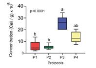
Spermatogonial cell concentration (cell/g) of Hyphessobrycon boulengeri. Spermatogonial cell concentration observed at protocols tested. P1(1.5 M methanol + 4.5 M propylene glycol), P2(1.5 M methanol + 5.5 M Me2SO), P3(2 M Me2SO + 2.5 M ethylene glycol) and P4(3M propylene glycol + 3 M Me2SO). Different letters indicate differences between the groups by the Kruskal-Wallis test followed by Dunn test.
The cellular viability was different (p<0.001), there was a difference among the P3 (25.42± 4.19%) and other treatments, P1 (5.84±4.32%), P2(5.19±1.15%) and P4 (13.16±4.8%) (Fig. 4).

Cellular viability (%) observed in the testicular tissue vitrified of Hyphessobrycon boulengeri with the protocols tested. Different letter indicated significantly differences by Tukey test.
Experiment 2
The cellular concentration was different (F (2, 24) = 32.53; p<0.0001) among the experimental groups, as it is showed in Fig. 5. There was a difference among all the recipients of cryopreservation tested. The highest cellular concentration of spermatogonial cells, was in a cryotube group, followed of the samples cryopreserved in gelatine capsules, while the samples cryopreserved at hypromellose capsules obtained the lowest cellular concentration.

Spermatogonial cell concentration (Cell/g) of Hyphessobrycon boulengeri, recovered in cryotubes, gelatin capsules and hypromellose capsules. Different letters indicate differences between groups using the ANOVA test followed by the Tukey test.
The cellular variability was different (F (2, 6) = 15.45; p=0.0043) between the experimental groups (Fig. 6). The greatest viability was observed in the samples cryopreserved in cryotubes (19.82±2.64%) and gelatin capsules (16.09±1.58%), those were significantly different to the hypromellose capsules with 8.74±3.01% of cellular viability.

Cellular viability (%) observed in the testicular tissue vitrified of Hyphessobrycon boulengeri sustained in cryotubes, gelatin capsules and hypromellose capsules. Different letters indicate significative difference using the Tukey test.
There was no difference (F (3, 20) = 0.9237; p=0.4474) among the experimental groups for the mitochondrial activity after cryopreservation (Fig. 7). The mitochondrial activity was varied of 12.30 to 18.41 AU/g, without difference among the groups.

Mitochondrial activity observed in the testicular tissue vitrified of Hyphessobrycon boulengeri sustained in cryotubes, gelatin capsules and Hypromellose capsules. Different letters indicate significative difference using the Tukey test.
The lipid peroxidation analyzed by the TBARS test was not different among the treatments (p=0.1308) presenting in Fig. 8.
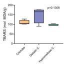
Lipid peroxidation by the analysis of the Thiobarbituric Acid Reactive Substance – TBARS (nmol of MDA/g) observed in the testicular tissue vitrified of Hyphessobrycon boulengeri sustained in cryotubes, gelatin capsules and Hypromellose capsules. Different letters indicate significative difference using the Tukey test.
A significant difference was observed (F (2.14) = 6.943; p=0.0080) for the antioxidative capacity measured by FRAP analysis, among the experimental groups (Fig. 9). The highest values observed were in samples cryopreserved in cryotubes (57.56±5.99 µg/Acid ascorbic equivalent) and the hypromellose capsules (55.42±9.8 µg/Acid ascorbic equivalent), which are significantly different from the gelatin capsule (39.94±9.06 µg acid ascorbic equivalent).

Antioxidant capacity measured with the method FRAP (Ferric Reducing Antioxidant Power) observed in the testicular tissue vitrified of Hyphessobrycon boulengeri sustained in cryotubes, gelatin capsules and Hypromellose capsules. Different letters indicate significative difference by the Tukey test.
DISCUSSION
Cryopreservation can maintain cells at exceptionally low temperatures with the intention to preserved their functionality (França et al. 2023). Vitrification it is considered a simple method since a sophisticated instrumental was no require, easy to do and their accessibility in different places. After that, the use of simple and functional artifacts is fundamental for the execution of vitrification. This article proposes and validates a protocol to vitrify fish testicular tissue in low-cost, biodegradable and easily accessible medical capsules in any country. And the results obtained here, in fact, glimpse a potential use of gelatin and hypromellose capsules to be used in the vitrification of fish testicular tissue, as a low-cost solution and conceptually aligned with the use of biodegradable material.
The execution of Experiment 1 in this study was essential to guarantee the best performance regarding the permeable cryoprotectant for vitrification of H. boulengeri testicular tissue. Even though, using the protocol by (Marques et al. 2018) as a reference in Piaractus mesopotamicus, the species-specific hypothesis was taken into account, both for the cryoprotectant and its concentration (Zidni et al. 2022). And the protocol by (Marques et al. 2018) was actually confirmed with DMSO as efficient for the testicular tissue of H. boulengeri.
The time that testicular tissue was exposure to the exposition/vitrification solution was significantly important for the action of the cryoprotectants to penetrate the tissue. Is demonstrated the less toxicity Lahnsteiner (2008) and high permeability of dimethilsufoxide (Seki et al. 2007) as cryoprotectant. On the other hand, ethylene glycol has the property of being hygroscopic and mixable with polar solvents (Rebsdat & Mayer 2012). According to (Lujić et al. 2017), the combination of two cryoprotectants in concentrations almost identical allows the general use of cryoprotectants in high concentrations, but in combination each one results in low quantity to be toxic for the cells. In this case, the exposure to (VS3) obtained a higher concentration of cells and viability after thawing using the trypan blue.
The results regarding the maintenance and viability of H. boulengeri sperm tissue after cryopreservation in capsules showed greater efficiency when the origin was gelatin instead of hypromellose. The concentration of sperm cells recovered after cryopreservation was much lower compared to cryotubes, the percentage of viability was equal to the traditional container used in cryopreservation protocols.
As for the better result for cell viability after vitrification of H. boulengeri testicular tissue observed in the gelatin capsule in relation to the hypromellose capsule, it may be related to its mechanical properties originating from proteins (Jongjareonrak et al. 2006). Another factor that can be considered was reported by (Cole et al. 2002) in studies with drug release. In this case, the authors related differential solubility and possible fragmentation of the polymer during enzymatic digestion. In recent studies, (França et al. 2023) showed that the functionality of sperm of Rhamdia quelen after cryopreservation was maintained and able to produce viable larvae. In the same way, was demonstrated that sperm quality of Mediterranean fishes was maintained in biodegradable capsules (hard-gelatin and hard-HPMC) (França et al. 2024).
Mitochondria are organelles with the responsibility to provide energy to the cells in the form of ATP (Kholodnyy et al. 2020). Eventually, the exposition to low temperatures can causes sensibility to mitochondria organelles (Tsvetkov & Naydenova 1987) and their validation functionality is crucial. In this study, the mitochondrial activity not vary from the other experimental groups, that indicated that cryotubes and biodegradables capsules corroborates their similarity as recipients to preserved the mitochondrial activity of the cells after warming. In addition to there, being no difference in the mitochondrial cell activity among the types of artifacts tested, the time of equilibrium and vitrification protocol used for ovarian tissue in P. mesopotamicus proposed by (Marques et al. 2018) was efficient. The results about cellular viability of the present study using acupuncture needles for vitrification were similar to those reported by (Marinovic et al. 2019) in spermatogonia of zebrafish.
The physiological disbalance between the production of reactive oxygen species (ROS) and antioxidant defenses can cause oxidative stress (West et al. 2007), resulting in DNA damage and the induction of lipid peroxidation, affecting membrane function, fluidity and structure (Freeman & Crapo 1982). For this study, TBARS levels be alike among treatments (p=0.1308) which indicates that the lipid peroxidation was not altered in the samples and the cryoprotectant used Me2SO provided a reduction in osmotic and mechanical stress as described by (Wang et al. 2007). Cellular antioxidant defense is mediated by mechanisms by which cells cancel the reactivity or inhibit the production of free radicals (Thornalley & Vasak 1985, Palamada & Kehrer 1992). The results showed that the samples cryopreserved in cryotubes and hypromellose capsule obtained higher values to the antioxidant potential determined by FRAP method in comparation with gelatin capsule.
Although we expected a greater antioxidant potential for cryopreservation using gelatin capsules, the lower antioxidant potential observed for this form of cryopreservation is not in itself a limiting factor for it is use. In fact, this reduction in antioxidant potential did not reflect on tissue oxidative damage, since there was no change in lipid peroxidation. Added to this, the results of cell viability, mitochondrial activity and spermatogonial cell concentration corroborate to highlight gelatin capsules as an excellent option for cryopreservation of testicular tissue of H. boulengeri.
In the present study, the cellular viability after vitrification/warming was demonstrated by the correct use of a protocol according to the fish species to optimize the cryopreservation of H. boulengeri testicular tissue. Also, it was proved that biodegradables capsules can be used as an eco-friendly container and low-cost alternative.
Acknowledgements
Coordenação de Aperfeiçoamento de Pessoal de Nível Superior (CAPES) [grant number 23038.011255/2021–17]. Danilo Pedro Streit Jr. (305387/2022-7), Thaíza Rodrigues de Freitas (141423/2021-8) are research fellows from the Conselho Nacional de Desenvolvimento Científico e Tecnológico (CNPq-Brazil). Additionally, we extend our appreciation to Energética Barra Grande S/A (BAESA) for their financial support [grant number P&D ANEEL 03936-1410/2021].
References
- ADAMOV NS ET AL. 2017. Triploid induction in the yellowtail tetra, Astyanax altiparanae, using temperature shock: Tools for conservation and Aquaculture. J World Aquac Soc 48: 741-750.
- BENZIE IF & STRAIN JJ. 1996. The ferric reducing ability of plasma (FRAP) as a measure of “antioxidant power”: the FRAP assay. Anal Biochem 239: 70-76.
- BRITSKY HA. 1972. Peixes de água doce de Estado de São Paulo: Sistemática. In: Comissão Interestadual da Bacia Paraná-Uruguai (Eds), Poluição e Piscicultura São Paulo, Faculdade de Saúde Pública da USP e instituto de Pesca, São Paulo, Brasil, p. 83-108.
- COELHO GCZ, YO IS, MIRA-LÓPEZ TM & YASUI GS. 2019. Preparation of a fish embryo for micromanipulation: staging of development, removal of the chorion and traceability of PGCs in Prochilodus lineatus Int J Dev Biol 63: 57-59.
- COLE ET, CONNOR AL, WILDING IR, PETEREIT HU, SCHMINKE C, BECKERT T & CADÉ D. 2022. Enteric coated HPMC capsules designed to achieve intestinal targeting. International J Pharm 231: 83-95.
- FRANÇA TS ET AL. 2023. Fish sperm cryopreservation using biodegradable containers: New low-cost and environment-friendly methodology. Reprod 166: 89-97.
- FRANÇA TS ET AL. 2024. Successful cryopreservation in biodegradable containers of sperm from aquaculture Mediterranean fishes. Theriogenology 216: 53-61.
- FREEMAN BA & CRAPO JD. 1982. Biology of disease. Free radicals and tissue injury. Lab inves 47: 412-426.
- HAGEDORN MM, DALY JP, CARTER VL, COLE KS, JAAFAR Z, LAGER CVA & PARENTI LR. 2018. Cryopreservation of Fish Spermatogonial Cells: The Future of Natural History Collections. Sci Rep 8: 6149.
- JONGJAREONRAK A, BENJAKUL S, VISESSANGUAN W, PRODPRAN T & TANAKA M. 2006. Characterization of edible films from skin gelatin of brownstripe red snapper and bigeye snapper. Food Hydrocoll 20: 492-501.
- KHOLODNYY V, BORYSHPOLETS S, DZYUBA B & COSSON J. 2020. Energetics of fish spermatozoa. In: Betsy J & Kumar S (Eds), Cryopreservation of Fish Gametes. Springer, Singapore, p. 69-116.
- LAHNSTEINER F. 2008. The effect of internal and external cryoprotectants on zebrafish (Danio rerio) embryos. Theriogenology 69: 384-396.
- LEE S, IWASAKI Y, SHIKINA S & YOSHIZAKI G. 2013. Generation of functional eggs and sperm from cryopreserved whole testes. PNAS 110: 1640-1645.
- LIU Y, PETERSON DA, KIMURA H & SCHUBERT D. 2002. Mechanism of cellular 3-(4,5- Dimethylthiazol-2-yl)-2,5-Diphenyltetrazolium Bromide (MTT) reduction, J Neurochem 69: 581-593.
- LUJIĆ J, MARINOVIĆ Z, SUŠNIK BAJEC S, DJURDJEVIČ I, KÁSA E, URBÁNYI B & HORVÁTH Á. 2017. First successful vitrification of salmonid ovarian tissue. Cryobiology 76: 154-157.
- MARINOVIĆ Z, LUJIĆ J, KÁSA E, CSENKI Z, URBÁNYI B & HORVÁTH Á. 2018. Cryopreservation of Zebrafish Spermatogonia by Whole Testes Needle Immersed Ultra-Rapid Cooling. JoVE 133: e56118.
- MARQUES L, FOSSATI AAN, LEAL MS, RODRIGUES RB, BOMBARDELLI RA & STREIT JR DP. 2018. Viability assessment of primary growth oocytes following ovarian tissue vitrification of neotropical teleost pacu (Piaractus brachypomus) Cryobiology 82: 118-123.
- MARQUES L, FOSSATI AAN, RODRIGUES RB, DA ROSA HT, IZAGUIRRY AP, RAMALHO JB, MOREIRA JCF, SANTOS FW, ZHANG T & STREIT JR DP. 2019. Slow freezing versus vitrification for the cryopreservation of zebrafish (Danio rerio) ovarian tissue. Sci Rep 9: 1-11.
- MATTHEWS M & VARGA ZM. 2012. Anesthesia and euthanasia in zebrafish. ILAR J 53: 192-204
- OHKAWA H, OHISHI N & YAGI K. 1979. Assay for lipid peroxides in animal tissues by thiobarbituric acid reaction. Anal Biochem 95: 351-358.
- PALAMADA J & KEHRER J. 1992. Inhibition of protein carbonyl formation and lipid peroxidation by glutathione in rat liver microsomes. Arch Biochem Biophys 293: 103-109.
- PIVA LH, DE SIQUEIRA-SILVA DH, GOMES CAG, FUJIMOTO T, SAITO T, DRAGONE LV, SENHORINI JA, PORTO-FORESTI F, FERRAZ JBS & YASUI GS. 2018. Triploid or hybrid tetra: Which is the ideal sterile host for surrogate technology? Theriogenology 108: 239-244.
- PORTO-FORESTI F, HASHIMOTO DT, SENHORINI JÁ & FORESTI F. 2010. Hibridação em piscicultura: monitoramento e perspectivas. In: Baldisserotto B & Gomes LC (Eds), Espécies nativas para piscicultura no Brasil, 2nd ed., Universidade Federal de Santa Maria – UFSM, Santa Maria, Brasil, p. 589-601.
- PŠENIČKA M, SITO T, RODINA M & DZYUBA B. 2016. Cryopreservation of early stage Siberian sturgeon Acipenser baerii germ cells, comparison of whole tissue and dissociated cells. Cryobiology 72: 119-122.
- REBSDAT S & MAYER D. 2012. Ethylene glycol. In: Ullmann’s Encyclopedia of Industrial Chemistry; Wiley-VCH Verlag Gmbh & Co, KGaA, Berlin, Germany, p. 552-546.
- SEKI S ET AL. 2013. Production of donor-derived offspring by allogenic transplantation of cryopreserved spermatogonia in medaka. Cryobiology 67: 398-442.
- SEKI S, KOUYA T, VALDEZ DM, JIN B, HARA T, SAIDA N, KASAI M & EDASHIGE K. 2007. The permeability to water and cryoprotectants of immature and mature oocytes in the zebrafish. Cryobiology 54: 121-124.
- THORNALLEY PJ & VASAK M. 1985. Possible role for metallothionein in protection against radiation-induced oxidative stress. Kinetics and mechanism of its reaction with superoxide an hydroxyl radicals. BBA 827: 36-44.
- TSVETKOV T & NAYDENOVA Z. 1987. Activity of ATP synthetase complex after low temperature treatment or freeze-drying of mitochondria isolated from skeletal muscles. Cryobiology 24: 280-284.
- WANG X, HUA TC, SUN DW, LIU B, YANG G & CAO Y. 2007. Cryopreservation of tissue-engineered dermal replacement in Me2SO: Toxicity study and effects of concentration and cooling rates on cell viability. Cryobiology 55: 60-65.
- WEST ER, XU M, WOODRUFF TK & SHEA LD. 2007. Physical properties of alginate hydrogels and their effects on in vitro follicle development. Biomaterials 28: 4439-4448.
- ZIDNI I, LEE HB, YOON JH, PARK JY, JANG HS, CHO YS, SEO YS & LIM HK. 2022. Cryopreservation of Roughscale Sole (Clidoderma asperrimum) Sperm: Effects of Cryoprotectant, Diluent, Dilution Ratio, and Thawing Temperature. Animals 12: 2553.


