Comparative analysis of the frequencies of α-thalassemia-associated mutations in microcytic patients and healthy volunteers in Rio de Janeiro, Brazil
Abstract
Thalassemia, an autosomal recessive genetic disorder affecting production of normal globin chains, presents varying prevalence worldwide. The frequency of global α-thalassemia remains understudied, especially in Brazil, due to its genetic diversity and large population size. This study compared α-thalassemia frequency between microcytic patients and healthy volunteers in Rio de Janeiro, stratified by self-reported skin color. DNA extracted from whole blood/EDTA underwent genotyping for α-thalassemia using multiplex PCR, targeting α3.7 kb, α4.2 kb, αSEA, αMED, αFIL, and α20.5 kb deletions. Frequency of α-thalassemia among patients was 63.6%, distributed as follows: 62.1% for α3.7 kb deletion, 0.7% for α4.2 kb deletion, and 0.8% for αSEA deletion. In healthy volunteers, the prevalence was 3.2%, distributed as 0.53% in self-declared white, 1.07% in brown, and 1.60% in black individuals, all presenting heterozygous genotype for α3.7 kb deletion. Frequency in healthy population was significantly lower than that observed in microcytosis population (p < 0.001). Determining α-thalassemia frequency in highly admixed population may enhance genetic counseling, given that asymptomatic carriers can produce offspring with combinations of Hb variants, potentially leading to severe clinical phenotypes.
Key words
admixed population; anemia; hemoglobinopathy; microcytosis; thalassemia
INTRODUCTION
Thalassemia is an autosomal recessive genetic disorder that disrupts the production of globin chains, leading to ineffective erythropoiesis, hemolysis, and anemia. Hemoglobin (Hb), composed of a heme group and four globin chains (two alpha and two non-alpha), is affected by this disorder. Although the global prevalence of thalassemias is not precisely known, it is estimated that 5% to 20% of the population carries mutations for alpha-thalassemia (α-thal) and around 1.5% for beta-thalassemia (β-thal). The incidence varies across different populations (Baird et al. 2022, Modell & Darlison 2008, Musallam et al. 2024, Vichinsky 2013).
For α-thalassemia, the production of Hb is reduced due to mutations affecting one, two, three, or all four alpha-globin genes (Musallam et al. 2024). The severity of the disease correlates with the number of functional genes impacted. Mutations in one gene typically cause α-thalassemia minor (α-thal+), leading to a mild or asymptomatic phenotype, often presenting as microcytosis, known as heterozygous α-thal+. Mutations in the α-globin genes can be clinically asymptomatic but may present with mild microcytosis and normal HbA/HbF proportions. These mutations can occur in two forms: (1) a heterozygous α⁰-thalassemia deletion (--/αα), where both genes are deleted on one chromosome, or (2) a homozygous α⁺-thalassemia (-α/-α), involving single-gene deletions on both chromosomes (Musallam et al., 2024). In cases where three genes are non-functional, the condition leads to Hb H disease, characterized by the formation of unstable Hb H, a tetramer of beta globins chains (β4), which have a high affinity for oxygen but are ineffective in oxygen delivery (Vichinsky 2013). The most severe form, α-thalassemia major (homozygous α⁰-thalassemia), results from the loss of all four α-globin genes (--/--), leading to hemoglobin Bart’s hydrops fetalis syndrome, a condition incompatible with extrauterine life (Musallam et al. 2024).
The mutations causing α-thalassemia can be either deletions or non-deletions. The most frequent α-thalassemia deletions worldwide are -α3.7 kb and -α4.2 kb, involving single α-globin gene losses on chromosome 16. In Southeast Asia, the -αSEA deletion (--αSEA/αα) predominates as a common α⁰-thalassemia variant, completely removing both α-globin genes while preserving embryonic ζ-globin genes. Homozygosity (--αSEA/--αSEA) results in α-thalassemia major (Hb Bart’s hydrops fetalis), a lethal condition (Vichinsky 2013). Non-deletion mutations (α-thal-αND) are often point mutations affecting key regulatory regions, leading to severe phenotypes similar to α-thal0 (Farashi & Najmabadi 2015). The clinical severity correlates directly with the degree of α-globin chain deficiency. A greater α:β globin chain imbalance results in more severe clinical manifestations. Notably, non-deletional α⁺-thalassemia variants often demonstrate more significantly reduced α-globin synthesis compared to deletional forms, leading to more pronounced clinical phenotypes despite being classified as α⁺-thalassemia (Farashi & Harteveld 2018, Vichinsky 2013).
Thalassemia prevalence is higher in regions such as Africa, India, the Mediterranean, the Middle East, and Southeast Asia. However, migration has altered the genetic landscape, impacting the frequency and distribution (Kattamis et al. 2020, Modell & Darlison 2008). Countries affected by hemoglobinopathies must integrate specific educational and diagnostic interventions into their healthcare systems and budgets to effectively address the needs of their populations (Piel & Weatherall 2014). There are significant gaps in knowledge about the global frequency of α-thalassemia, exacerbated by underreporting and the lack of molecular diagnostics (Musallam et al. 2024). This knowledge gap may hinder the prioritization of diagnostic interventions for α-thalassemia within the public health system (Piel & Weatherall 2014). Brazil’s population is highly admixed due to successive waves of migration, including the indigenous Amerindians, African slaves, and European immigrants. This genetic diversity complicates the determination of thalassemia mutation profiles in the country. Moreover, the vast territorial extension of Brazil contributes to the lack of consistent data on the prevalence of hemoglobinopathies, particularly α-thalassemia, which is harder to diagnose than β-thalassemia due to the need for complex techniques as DNA mutation analysis (Cardoso et al. 2012, Silva Filho et al. 2010, Fonseca et al. 2013, Wagner et al. 2010)
This study aimed to compare the frequency of α-thalassemia in two population groups in Rio de Janeiro, Brazil: patients with microcytosis seeking thalassemia diagnosis at the Clinical Analysis Laboratory of the Faculty of Pharmacy/UFRJ (LACFar/UFRJ) and healthy blood donors without clinical or laboratory complaints from a tertiary hospital. The findings highlight the need for better screening and diagnostic strategies to manage and understand the impact of α-thalassemia in such a genetically diverse population.
MATERIALS AND METHODS
Population studied
This cross-sectional cohort study involved two groups. The first included 998 patients with microcytosis complaints, referred by hematologists to LACFar/UFRJ between April 2009 and December 2023 for diagnostic evaluation. All patients who underwent diagnosis of α-thal during this period were included in the study. The control group consisted of 187 hematologically healthy volunteers randomly recruited from the hospital blood donor bank between March and December 2017 during routine donation visits. All donors from that period were invited to participate in the study but not all accepted. All participants completed a demographic questionnaire and self-classified their skin color according to the standardized Brazilian census categories (IBGE 2023), with the following distribution: 84 White, 53 Brown, and 50 Black individuals. The study was approved by the respective Research Ethics Committees, and all participants provided informed consent. Number of approval opinions from the CEP: CEP/INTO: 1.467.071 and CEP/HUCFF: 5.737.364.
Hematological and biochemical analysis and diagnosis of alpha-thalassemia
For patients attended at LACFar/UFRJ, a complete blood count (CBC) was performed using the Pentra ES 60 automated counter (Horiba ABX, France). Additionally, serum iron, latent iron binding capacity (LIBC), total iron binding capacity (TIBC), and transferrin saturation index (TSI) tests were carried out according to the manufacturer’s instructions on Labmax Plenno colorimetric biochemical analyzer (Labtest Diagnóstica/Diconex, Argentina). The transferrin saturation index (TSI) was used to assess the presence of iron deficiency anemia in the population with microcytosis, in accordance with the guidelines of the Ministry of Health (Brasil 2023), as this provides a more accurate analysis of iron metabolism as a whole. Ferritin was not used because it is an acute phase protein, with elevated serum concentrations in cases of infection, inflammation or neoplastic diseases. Serum iron was not used because some individuals use medicinal iron supplements, which can mask their serum concentrations, which remain close to normal without adequate monitoring of ferritin levels (Brasil 2023, Figueiredo 2010, Grotto 2010).
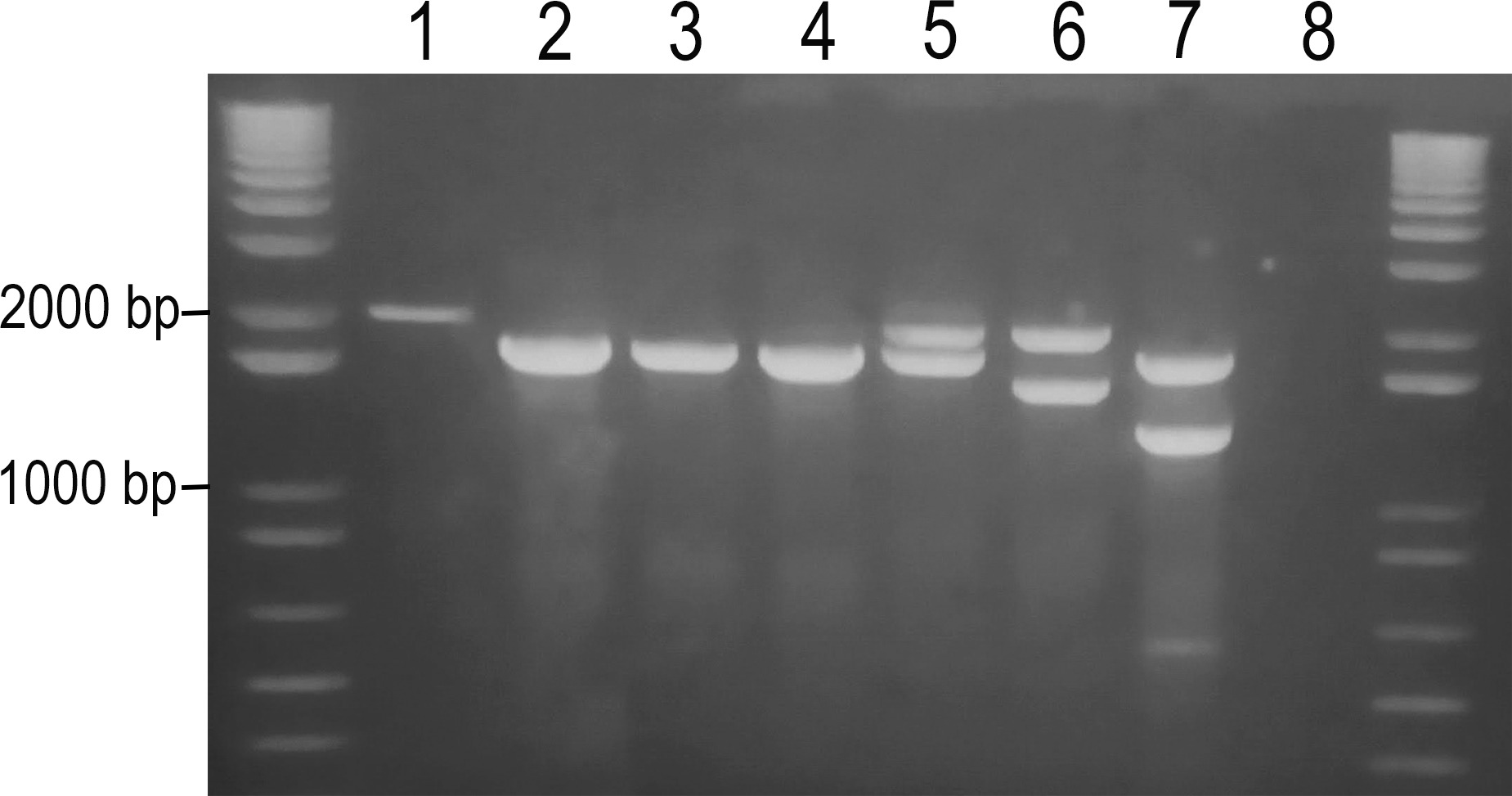
DNA was extracted from whole blood with EDTA using the Biopur Mini Spin Plus kit (Mobius - reference BP100-50) according to the manufacturer’s protocol. In both, patients and healthy volunteers, genotyping for α-thal was performed by multiplex polymerase chain reaction (PCR), following the method described by Chong et al. (2000). Most frequently found deletion mutations for α-thal were investigated, including α3.7 kb, α4.2 kb, αSEA, αMED, αFIL, and α20.5 kb deletions. Participants with a mutation in a single DNA strand (allele) were classified as heterozygous, while those with the same mutation in both alleles were classified as homozygous. Participants with different mutations in each DNA allele were classified as double heterozygous, and those without any of the studied mutations were classified as wild-type (WT) (Figure 1). In healthy volunteers, genotyping was performed blindly to hematological indices to prevent bias in the analysis.

Representative gel electrophoresis analysis of Multiplex PCR for α-thal screening. 1: Homozygous genotype for α3.7 kb deletion (2000 kb); 2, 3, 4: No mutations detected, WT (1800 kb); 5: Heterozygous genotype for α3.7 kb deletion (2000 kb/1800 kb); 6: Double heterozygous genotype for α3.7 kb/4.2 kb deletions (2000 kb/1600 kb); 7: Heterozygous genotype for αSEA deletion (1800 kb/1349 kb); 8: Negative control (water). Molecular weight markers are shown at both ends of the gel.
Weighted mean corpuscular volume (wMCV) was utilized to evaluate and compare microcytosis in patients, considering the significant age variation within this population. Weighting by age was implemented to normalize the value used, given the specific reference values for each age group (Siqueira et al. 2022).
Statistical analysis
A chi-squared test was performed to analyse the differences in frequency between the two populations (http://vassarstats.net/). Hematological data were analysed using descriptive statistics. Intergroup comparisons were conducted using GraphPad Prism v8.0.2 (GraphPad Inc., San Diego, California, USA). Patients were categorised based on their genotype. Groups containing seven or more individuals were analysed using a one-way ANOVA test with a Tukey’s multiple comparisons test to compare differences in continuous variables between all groups. A statistically significant difference was considered for results with p < 0.05. No statistical analysis was conducted for healthy volunteers.
RESULTS
Two groups of volunteers were selected to evaluate the frequency of α-thal mutations. In the healthy volunteers’ group, samples from 117 (62.6%) male and 70 (37.4%) female individuals were analyzed. The mean age was 34.5 years, with a mode of 39.0 years, and ages ranging from 16 to 74 years. Regarding educational levels, 11.8% had primary education, 57.2% had secondary education, 29.4% had higher education, and 1.6% had postgraduate education. In addition, a total of 998 patients at LACFar/UFRJ underwent the diagnostic protocol for α-thal. This population had a mean age of 16.9 years, with a mode of 2.0 years, and ages ranging from 4 months to 94 years. Among these patients, 48.8% were male and 51.2% were female. The level of educational attainment and skin color, whether self-identified or identified by others, were not evaluated in this population (Table I).
In the healthy volunteers group, we found six individuals carrying the heterozygous genotype for α3.7 kb deletion, representing 3.2% of this population, including one white woman, one brown man, one brown woman, and 3 women with black skin color, as presented in Table II.
Among the patients tested for α-thal from LACFar/UFRJ, 634 (63.5%) were positive for at least one of the mutations analyzed, while 364 (36.5%) did not exhibit any of the mutations studied. The following genotypic distribution for patients was observed: 33.0% were heterozygous for α3.7 kb deletion, 29.2% were homozygous for α3.7 kb deletion, 0.7% were heterozygous for αSEA deletion, 0.3% were double heterozygous for α3.7 kb/4.2 kb deletions, 0.2% were heterozygous for α4.2 kb deletion, 0.1% were homozygous for α4.2 kb deletion, and 0.1% were heterozygous for α3.7 kb/SEA deletions (Table II).
Furthermore, laboratory tests to evaluate anemia and iron deficiency were carried out only for LACFar/UFRJ patients. Regarding the hematimetric indices of this population, the following values were observed: slightly elevated red blood cell count (mean RBC 5.11 x 10^12/L + 0.68), mild anemia (mean Hb 11.7 g/dL +1.5), significant microcytosis (mean wMCV 66.0 fL + 6.1) and hypochromia (mean MCH 23.0 pg + 2.8), with iron metabolism within the reference range (mean TSI 25.6% + 11.1; serum iron 80.6 μg/dL + 34.6; TIBC 319.8 μg/dL + 58.0) (Table II).
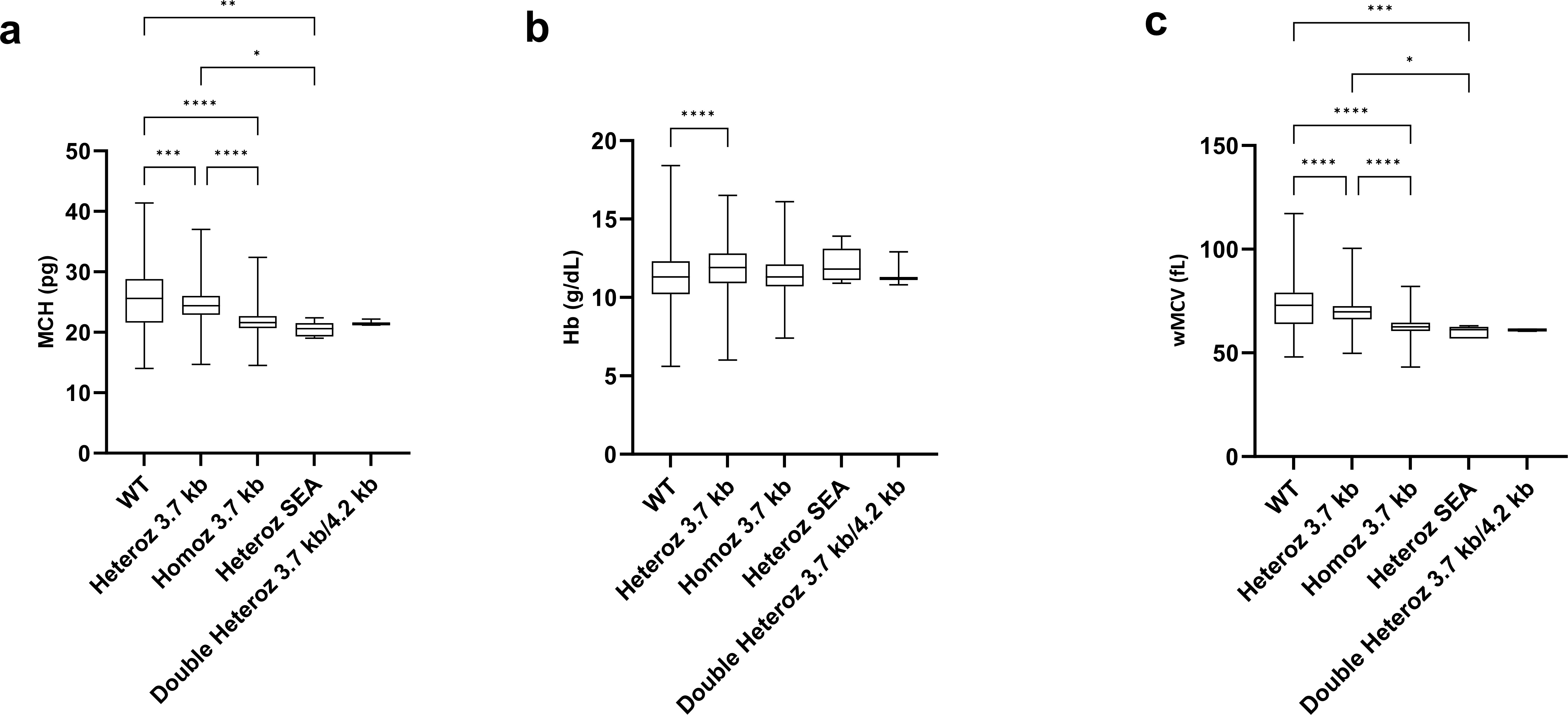
A highly significant association between microcytosis and α-thalassemia was observed (Chi-Square test, p < 0.0001), indicating individuals with microcytosis had substantially higher probability of carrying α-thalassemia genotypes compared to controls. Statistical analysis of LACFar/UFRJ patient data revealed significant differences in three key hematological parameters: weighted mean corpuscular volume (wMCV), mean corpuscular hemoglobin (MCH), and hemoglobin concentration (Hb) (Figure 2).

Comparison of hematological parameters among α-thalassemia genotypes. (a) Mean corpuscular hemoglobin (MCH) levels, (b) Mean hemoglobin (Hb) concentration and (c) Weighted mean corpuscular volume (wMCV) for each identified genotype. *: p < 0.05; **: p < 0.01; ***: p < 0.001; ****: p < 0.0001.
Comparative statistical analysis revealed a significant difference in Hb, wMCV, and MCH between the WT and Homozygous α3.7 kb groups, compared to other genotypes. Additionally, Heterozygous α3.7 kb x Homozygous α3.7 kb exhibited a difference in wMCV and MCH, and a statistical difference was observed in wMCV between the genotypes WT x Heterozygous αSEA and Heterozygous α3.7 kb x Heterozygous αSEA. Regarding the number of RBC and TSI, no statistical difference was found between the genotype groups (data not shown).
DISCUSSION
Thalassemias are genetic disorders that significantly impact individuals’ quality of life, potentially leading to severe anemia and recurrent blood transfusions. Despite their serious implications, there is a lack of comprehensive studies on the frequency of mutations in unbiased populations, particularly in Brazil. This study analyzed two groups: patients referred for hemoglobinopathy diagnoses, who are likely biased due to symptomatic presentation, and a healthy volunteer population. The high α-thalassemia frequency observed in the patient group does not reflect the true prevalence in Rio de Janeiro. Although many carriers are asymptomatic, the genetic nature of α-thal and the limited genetic counseling services in Brazil mean that the number of individuals affected could be higher if asymptomatic carriers have children together (Brunoni 2002). Each asymptomatic heterozygous couple has a 25% chance of having a child with a severe form of the disease. Chi Square test revealed a significant difference in the risk of positive α-thal genotypes between the two populations, highlighting the diagnostic bias in the patient group compared to the healthy volunteers.
The most significant statistical difference in genotype frequencies was observed between the heterozygous α3.7 kb group and the wild-type (WT) group (Figure 2). The WT group, used for comparison, included patients referred to the laboratory who lacked the mutations under investigation but exhibited notable microcytosis and hypochromia, with mean values of wMCV = 72.2 fL and MCH = 25.4 pg.
The α3.7 kb deletion is the most frequent mutation among the two populations studied, found in 98% of LACFar/UFRJ patients positive for α-thal. Indeed, the frequency of this mutation, commonly observed in African and Mediterranean populations, mirrors its prominence in our study population (Wagner et al. 2010). This observation underscores the genetic diversity present in our cohort and highlights the importance of considering regional genetic patterns when assessing thalassemia frequency and associated risks. Other authors found the α3.7 kb deletion to be the most frequent in their populations, corroborating our result (Anselmo et al. 2021, Souza et al. 2015). Souza et al. (2015) investigated thalassemia frequency in two populations; revealing rates of 12.8% among newborns and 14.9% among blood donors disqualified due to low hematocrit or anemia. Similarly, Anselmo et al. (2021) reported a frequency of 8.0%.
The next most frequent mutation identified in this study was the heterozygous αSEA genotype, present in 7% of patients. There are no other records of the frequency of this mutation in the Brazilian population for comparison, although other authors have researched it. As far as we know, only Siqueira et al. (2022) published two patients with this mutation in the Brazilian population using part of our database (Anselmo et al. 2021, Souza et al. 2015, Wagner et al. 2010). Ruengdit et al. (2023) reported that this mutation has significant frequency in Southeast Asian countries, ranging from 4% to 14%. Although there are no references to significant immigration from Southeast Asian countries to Brazil, Oliveira and Masiero highlight significant political and economic collaborations between Brazil and Asian countries, which generally attract migratory movements between the involved countries. This may explain the presence of this genotype in the studied population (Oliveira & Masiero 2005). This mutation was also observed in double heterozygosity with the α3.7 kb deletion in only one patient of Asian origin, who presented typical thalassemia clinical phenotype: significant microcytosis (wMCV = 50.4 fL) and hypochromia (MCH = 15.9 pg), elevated RBC (6.02 x 10^12/L), anemia (Hb = 9.6 g/dL), and no iron deficiency (TSI = 33.2%). According to Vichinsky (2013), αSEA deletion has a slightly more severe phenotype than the α3.7 kb deletion, as it is an α-thal0 mutation, while the α3.7 kb is an α-thal+. The α4.2 kb deletion was found in simple heterozygosity (0.2%), double heterozygosity with the α3.7 kb deletion (0.3%), and homozygosity (0.2%). This mutation is commonly found in Asian and Mediterranean populations (Wagner et al. 2010). In the Brazilian population, two studies have identified it. Souza et al. (2015) found three (0.35%) newborns with a heterozygous α4.2 kb genotype and African ancestry, and Anselmo et al (2020) found one (0.1%) blood donor with no information about his ancestry. All three genotypes identified in the studied population exhibited significant microcytosis and hypochromia (Anselmo et al. 2020, Souza et al. 2015). In addition to these, other authors also researched this mutation and did not find it (Anselmo et al. 2020, Silva Filho et al. 2010, Souza et al. 2015, Wagner et al. 2010).
In the comparative analysis of genotype groups, wMCV was the parameter with the greatest statistical difference (Figure 2). Microcytosis, a key feature of thalassemia, is associated with reduced wMCV. As anticipated, the WT group exhibited significant differences in wMCV compared to all other genotypes, and the heterozygous α3.7 kb genotype showed differences from the homozygous α3.7 kb genotype. The homozygous genotype had a lower wMCV value, which was expected, since it has the deletion of the α-globin gene in both alleles and a consequent reduction in the synthesis of the α-chain. This genotype-phenotype correlation aligns with the expected more severe hematological manifestation in homozygous individuals compared to heterozygous carriers. No significant differences were found between the heterozygous αSEA and homozygous α3.7 kb genotypes for any parameter. This aligns with Vichinsky’s (2013) prediction that the αSEA deletion, associated with the α-thal0 phenotype, is more severe than the α3.7 kb deletion, which is associated with the α-thal+ phenotype. Note that a single allele with the αSEA deletion is capable of presenting a phenotype similar to two alleles with the α3.7 kb deletion, by the length of the deleted DNA fragment. This will show a phenotype with greater clinical severity (Farashi & Harteveld 2018, Vichinsky 2013).
The second population investigated in this study was composed of healthy volunteers. The gender distribution among healthy volunteers was also consistent with data from Lisot & Silla (2004), who investigated the prevalence of hemoglobinopathies in blood donors in Rio Grande do Sul state, finding 62% male and 38% female. Our data are also consistent with the 9th Hemotherapy Production Report of Brazil (ANVISA 2022), which found that in 2020, 56% of Brazilian donors were male and 44% were female.
The frequency found in this group was significantly different from that of patients from LACFar/UFRJ, as expected. Genotyping of healthy individuals aimed to determine the frequency of α-thal in the healthy population of the state of Rio de Janeiro without the bias present in the patients attended at LACFar/UFRJ. As previously discussed, identifying asymptomatic carriers of thalassemias in the healthy population is important due to the high miscegenation in the Brazilian population. These asymptomatic adult carriers likely have not and will not experience any clinical complications due to α-thal; however, individuals who inherit combinations of Hb variants, such as Hb S, C, E, or β-thal with α-thal, can present severe clinical phenotype. These asymptomatic carriers can produce offspring with these combinations of Hb variants along with α-thal, thereby increasing the frequency of symptomatic carriers with the potential for a severe phenotype in our population. Compared to other studies on the Brazilian population, few studies besides this one have conducted genotyping for α-thal in healthy blood donors (Anselmo et al. 2020, Lisot & Silla 2004, Sonati et al. 1991) and none of these referenced observations by self-declared skin color. The study by Sonati & Costa (1990) examined a population of 47 Black donors in São Paulo state. No hematimetric data of the participants were recorded; the authors found 29.8% of the population with α-thal, investigating only the α3.7 kb deletion using restriction enzymes. Lisot & Silla (2004) investigated the presence of Hb H in 608 participants, finding Hb H in only 0.66% of the population; they used heteroidentification, evaluated hematimetric indices, and employed Hb H testing as their methodology. There is no mention in Anselmo et al. (2020) if the sampling was made blindly to the hematimetric indices, but they found a slightly higher frequency of α-thal (5.5%) in comparison to the present study. However, the population studied by Anselmo et al. (2020), consisting of 989 blood donors, was primarily composed of individuals from the Amazon Region with a strong Amerindian genetic heritage. This genetic background may have influenced the differences in frequencies found compared to our study.
This study is the first to use self-declared skin color data for investigating α-thal in Brazil, since Lisot & Silla (2004) evaluated the ethnic group of participants using heteroidentification. These authors used Hb H testing for diagnosing α-thal, not a molecular methodology, which leads to the underreporting of milder cases since one or two non-functional α-genes can be present without Hb H. They found 4/608 (0.66%) participants positive for α-thal, with 3/437 (0.67%) of European descent and 1/157 (0.64%) of mixed descent (Lisot & Silla 2004). Molecular methodology is more specific for diagnosing α-thal, as it can identify deletions of just one or two of four α-globin genes. Hb H consists of β-chain tetramer (β4), whereas normal Hb formation requires two α and two β chains (α2β2). Hb H occurs only when three or four α-globin genes lose their function. Given the complexity required for Hb H formation, it is suggested that asymptomatic carriers (with two or three functioning α-genes) are unlikely to be identified by this technique. In the present study, employing molecular techniques, 6/187 (3.2%) participants from the healthy donor cohort were detected as heterozygous for α3.7 kb deletion. Had Hb H testing been employed for diagnosis, none would have been identified with a positive α-thal genotype. Lisot & Silla (2004) reported a frequency nearly five times lower (0.66%) despite a sample size approximately three times larger (4 out of 608). This discrepancy could be attributed to the variation in methodologies utilized.
In the healthy volunteers population, only α3.7 kb deletion was found, which is the most common mutation worldwide and in the Brazilian population, as previously discussed.
When comparing our findings with other studies on α-thal frequency in the Brazilian population, α3.7 kb deletion emerges as the most investigated mutation, as previously highlighted. Even investigations targeting different α-thal mutations frequently encounter the α3.7 kb deletion. Other research groups have also focused on biased populations. For instance, Souza et al. (2009) studied patients with microcytosis and anemia, revealing a 9.1% α-thal frequency; Cardoso & Guerreiro (2010) examined individuals with Hb S, reporting a 16.2% α-thal frequency; and Souza et al. (2015) investigated donors disqualified due to low hematocrit, finding a 14.9% α-thal frequency. Among these, Souza et al. (2009) identified the highest frequency, reporting a 20.4% frequency of α-thal in patients with microcytosis and hypochromia but no anemia. The significant difference between our findings and those of other studies may be attributed to the fact that our patients are referred to LACFar/UFRJ for diagnosis based on existing hematological complaints or symptoms, unlike the routine hospital patient cohorts examined in other studies.
These results highlight the importance of screening for α-thalassemia in individuals who present microcytosis, with or without anemia. Since α-thalassemia is rarely considered, these individuals may unnecessarily take iron supplements without achieving an adequate therapeutic response, potentially leading to harm. Population-based screening for α-thalassemia should be implemented, even in asymptomatic individuals, in order to enhance genetic counseling. This approach is particularly valuable since many carriers are clinically silent, and molecular testing provides superior accuracy compared to conventional methods.
While our findings are based on laboratory diagnostic data without clinical follow-up, they provide important baseline information about α-thalassemia distribution patterns. This study makes a significant contribution as the first to compare hematological parameters between diagnosed patients and a healthy control population stratified by self-reported skin color using standardized Brazilian census categories. These results help address the notable lack of population-level data on α-thalassemia in our region and nationally, offering valuable insights for future research and potential screening programs.
CONCLUSIONS
The frequency of α-thal was evaluated in two populations from the state of Rio de Janeiro. Among patients with microcytosis who sought the LACFar/UFRJ for the diagnosis of thalassemias/hemoglobinopathies, a frequency of 63.6% was found, distributed as follows: 62.1% for α3.7 kb, 0.7% for α4.2 kb and 0.8% for αSEA deletions. In healthy individuals (blood donors), frequence of 3.2% was found, distributed as follows: 0.53% in individuals self-identified as white, 1.07% as brown, and 1.60% as black; all with the heterozygous genotype for α3.7 kb deletion. Our findings demonstrate distinct α-thalassemia detection patterns between the studied populations. In the healthy control group, without hematological complaints, we observed a significantly lower α-thalassemia frequency compared to the clinically referred population presenting with marked microcytosis. This expected pattern reflects fundamental principles of diagnostic epidemiology, where targeted testing of symptomatic or high-risk individuals naturally yields higher detection rates than general population screening. The magnitude of this difference underscores the importance of clinical context when interpreting α-thalassemia prevalence data
Acknowledgements
The authors would like to thank all the professionals on the LACFar team and the interns involved in this work for their participation, as well as HORIBA Instruments Brazil for their support. The study was funded by Fundação Carlos Chagas Filho de Amparo à Pesquisa do Estado do Rio de Janeiro – FAPERJ (E-26/210.925/2024).
References
- AIRD DC, BATTEN SH & SPARKS SK. 2022. Alpha- and Beta-thalassemia: Rapid Evidence Review. Am Fam Physician 105(3): 272-280.
- ANSELMO FC, FERREIRA NS, MOTA AJ, GONÇALVES MDS, ALBUQUERQUE SRL, FRAIJI NA, FERREIRA ACD & MOURA NETO JP. 2020. Deletional Alpha-Thalassemia Alleles in Amazon Blood Donors. Adv Hermatol 1: 4170259.
- ANSELMO FC, SOUMANOU AG, AGUIAR FERREIRA C, SOBRINHA FMV, CASTRO ACS, BRITO RO, MOTA AJ, SOUZA GONÇALVES M & MOURA NETO JP. 2021. Detection of common deletional of α-thalassemia 3.7 kb from metropolitan region of manaus, Amazonas, Brazil. Mediterr J Hermatol Infect Dis 13(1): 1-8.
-
ANVISA - AGÊNCIA NACIONAL DE VIGILÂNCIA SANITÁRIA. 2024. 9th Hemotherapy Production Report of Brazil. Available at: https://app.powerbi.com/view?r=eyJrIjoiMWM4MDQzNDMtYjZjZC00ZTBhLWFkOTctODdiZjE2ODQ4YTJkIiwidCI6ImI2N2FmMjNmL WMzZjMtNGQzNS04MGM3LWI3MDg1ZjVlZG Q4MSJ9. Accessed May 6, 2024.
» https://app.powerbi.com/view?r=eyJrIjoiMWM4MDQzNDMtYjZjZC00ZTBhLWFkOTctODdiZjE2ODQ4YTJkIiwidCI6ImI2N2FmMjNmL -
BRASIL. 2023. Protocolo Clínico e Diretrizes Terapêuticas - Anemia por Deficiência de Ferro. Available at: https://www.gov.br/conitec/pt-br Accessed May 6, 2024.
» https://www.gov.br/conitec/pt-br -
IBGE - INSTITUTO BRASILEIRO DE GEOGRAFIA E ESTATÍSTICA. 2023. Censo demográfico 2022: identificação étnico-racial da população, por sexo e idade: resultados do universo. Available at: https://biblioteca.ibge.gov.br/index.php/biblioteca-catalogo?view=detalhes&id=73105. Accessed June 2, 2025.
» https://biblioteca.ibge.gov.br/index.php/biblioteca-catalogo?view=detalhes&id=73105 -
BRUNONI D. 2002. Genetic Counseling. DOI: https://doi.org/10.1590/S1413-81232002000100009
» https://doi.org/10.1590/S1413-81232002000100009 - CARDOSO GDL & GUERREIRO JF. 2010. Molecular characterization of sickle cell anemia in the Northern Brazilian state of Pará. Am J Hum Biol 22(5): 573-577.
- CARDOSO GL, TAKANASHI SYL & GUERREIRO JF. 2012. Inherited hemoglobin disorders in an Afro-Amazonian community: Saracura. Genet Mol Biol 35(3): 553-556.
- CHONG SS, BOEHM CD, HIGGS DR & CUTTING GR. 2000. Single-tube multiplex-PCR screen for common deletional determinants of α-thalassemia. Blood 95(1): 360-362.
- FARASHI S & HARTEVELD CL. 2018. Molecular basis of α-thalassemia. Blood Cells Mol Dis 70: 1-9.
- FARASHI S & NAJMABADI H. 2015. Diagnostic pitfalls of less well recognized HbH disease. Blood Cells Mol Dis 55(4): 1-6.
- FIGUEIREDO MS. 2010. Impacto da inflamação na regulação do ferro e deficiência funcional de ferro. Rev Bras Hematol Hemoter 32(supl. 2): 22-28.
- FONSECA SF, MOURA NETO JP & GONÇALVES MS. 2013. Prevalence and molecular characterization of β-Thalassemia in the state of Bahia, Brazil: First identification of mutation HBB: C.135delC in Brazil. Hemoglobin 37(3): 285-290.
- GROTTO HZW. 2010. Diagnóstico laboratorial da deficiência de ferro. Rev Bras Hematol Hemoter 32(supl. 2): 9-11.
- KATTAMIS A, FORNI GL, AYDINOK Y & VIPRAKASIT V. 2020. Changing patterns in the epidemiology of β-thalassemia. Eur J Haematol 105(6): 692-703.
- LISOT CLA & SILLA LMDR. 2004. Screening for hemoglobinopathies in blood donors from Caxias do Sul, Rio Grande do Sul, Brazil: prevalence in an Italian colony. Cad Saude Publica 20(6): 1595-1601.
- MODELL B & DARLISON M. 2008. Global epidemiology of haemoglobin disorders. Bull World Health Organ 86(6): 480-487.
- MUSALLAM KM, CAPPELLINI MD, COATES TD, KUO KHM, AL-SAMKARI H, SHETH S, VIPRAKASIT V & TAHER AT. 2024. Αlpha-thalassemia: A practical overview. Blood Rev 64: 1001-1015.
- OLIVEIRA HA & MASIERO G. 2005. Estudos Asiáticos no Brasil: contexto e desafios. Rev Bras Polit Int 48: 5-28.
- PIEL FB & WEATHERALL DJ. 2014. The α-Thalassemias. N Engl J Med 371(20): 1908-1916.
- RUENGDIT C, PUNYAMUNG M, INTASAI N & PORNPRASERT S. 2023. Single-tube multiplex real-time PCR with EvaGreen and high-resolution melting analysis for diagnosis of α0-thalassemia -SEA, -THAI, and -CR type deletions. PLoS One 18(11): 4-11.
- SILVA FILHO IL, RIBEIRO GS & PIMENTA-BUENO LM & SERPA MJA. 2010. The frequency of β-globin gene haplotypes, α-thalassemia and genetic polymorphisms in children with sickle cell disease in Rio de Janeiro, Brazil. Rev Bras Hematol Hemoter 32(1): 76-78.
- SIQUEIRA PFR, FLEURY MK, PONTES RM, SILVA RSP, COSTA ES & LAND MGP. 2022. Novel Decision Tool for More Severe α-Thalassemia Genotypes Screening with Functional Loss of Two or More α-Globin Genes: A Diagnostic Test Study. Diagnostics 12(12): 1-10.
- SONATI M & COSTA F. 1990. Hemoglobin Bart’s in a brazilian black population. Braz J Med Biol Res 23: 395-396.
- SONATI MF, FARAH SB, RAMALHO AS & COSTA FF. 1991. High prevalence of α-thalassemia in a black population of Brazil. Hemoglobin 15(4): 309-311.
- SOUZA AES, CARDOSO GL, TAKANASHI SYL & GUERREIRO JF. 2009. α-Thalassemia (3.7 kb deletion) in a population from the Brazilian Amazon region: Santarém, Pará State. Genet Mol Res 8(2): 477-481.
- SOUZA RAV, CARLOS AM, SOUZA BMB, RODRIGUES CV, ARAUJO PEREIRA G & MORAES-SOUZA H. 2015. α-thalassemia: genotypic profile associated with ethnicity and hematological differentiation of iron deficiency anemia in the Region of Uberaba, Minas Gerais, Brazil. Hemoglobin 39(4): 264-269.
- VICHINSKY EP. 2013. Clinical manifestations of α-Thalassemia. Cold Spring Harb Perspect Biol 5(5): a011742.
- WAGNER SC, CASTRO SM, GONZALEZ TP, SANTIN AP, FILIPPON L, ZALESKI CF, AZEVEDO LA, AMORIN B, CALLEGARI-JACQUES SM & HUTZ MH. 2010. Prevalence of common α-thalassemia determinants in south Brazil: Importance for the diagnosis of microcytic anemia. Genet Mol Biol 33(4): 641-645.