
 Prognostic significance and therapeutic outcome prediction through serum miR-21 and miR-145 in ovarian cancer
Abstract
This study investigated the diagnostic value of serum microRNA-21 (miR-21) and miR-145 in assessing the treatment response and prognosis of ovarian cancer patients. A total of 139 patients enrolled in this study were categorized based on their chemotherapy outcomes into three groups: partial response, stable disease, and disease progression. Significant differences in serum levels of miR-21 and miR-145 among these groups were observed. Specifically, elevated miR-21 and reduced miR-145 levels were associated with worse treatment outcomes and poorer prognoses. Multivariate logistic regression analysis identified miR-21 and miR-145, along with age, tumor stage, and lymph node metastasis, as independent predictors of patient outcomes. The receiver operating curve (ROC) analysis indicated that the combination of miR-21 and miR-145 provided higher accuracy in predicting patient prognosis compared to each biomarker alone. The findings suggest that miR-21 and miR-145 can be effective supplementary tools for evaluating treatment effectiveness and prognostic assessments in ovarian cancer patients.
Key words
miR-21; miR-145; ovarian cancer; chemotherapy; prognostic assessment
INTRODUCTION
Ovarian cancer is a highly malignant female reproductive organ tumor with high morbidity and mortality (Elies et al. 2018, González-Martín et al. 2024). It usually remains asymptomatic in the early stages, leading to a late diagnosis of the disease at mid-to-late stages when surgical intervention becomes impossible (Fujiwara et al. 2021, Harter et al. 2022, Lim et al. 2022). At the intermediate and advanced stages of ovarian cancer, chemotherapy becomes the primary intervention option that significantly improved the overall survival of patients with ovarian cancer.
The effectiveness of chemotherapy significantly influences the clinical prognostic assessment of patients (Mizuno et al. 2020, Park et al. 2023). Therefore, effective evaluation of the disease outcome and timely adjustment of treatment procedure play vital roles in improving the clinical anti-cancer efficacy and outcome. Approximately 50% of microRNAs (miRNAs), which have the capability to modulate the expression of multiple target genes, influencing various biological behaviors such as tumor proliferation, apoptosis and differentiation, are located in genomic regions associated with cancer (Chen et al. 2022, Syeda et al. 2020). Abnormal expression of miR-21 and miR-145 in multiple solid tumors including liver cancer and lung cancer have been reported (Arghiani & Matin 2021, Xu et al. 2020) and have been used as prognostic biomarkers for various cancers (Bautista-Sánchez et al. 2020, Jiang et al. 2020, Jin et al. 2019, Yang et al. 2014). MiR-21 has been utilized as diagnostic biomarker for ovarian cancer (Talaat et al 2022, Ralser et al. 2022), however, the prognostic value of miR-21 and miR-145 in ovarian cancer patients receiving chemotherapy remains unexplored.
In the current study we aim to analyze and explore the predictive value of serum miR-21 and miR-145 in the efficacy and outcomes of ovarian patients receiving standard chemotherapy.
MATERIALS AND METHODS
Basic information
This study included 139 ovarian cancer patients treated at our hospital from July 2018 to July 2020. The average age of the participants was 56.64±9.14. The average body mass index (BMI) was 23.58±1.64 kg/m2. Among these patients, 39 were postmenopausal and 100 were premenopausal. Regarding tumor differentiation, 60 cases were classified as poorly differentiated, while 79 cases were moderately to highly differentiated. In terms of pathology, 85 cases were diagnosed with serous ovarian cancer, and 54 cases with mucinous ovarian cancer. Lymph node metastasis was present in 30 cases, while 109 cases did not have lymph node metastasis. Additionally, 26 patients presented with refractory ascites, whereas 113 did not.
The study was approved by the ethics committee of the 10th affiliated hospital of Southern Medical University (Approval number: 20210001). Informed consent was obtained from each participant.
Inclusion and exclusion criteria
Inclusion criteria: (1) All participants met the diagnostic criteria for ovarian cancer, underwent pathological examinations, and were confirmed to have ovarian cancer; (2) All participants were newly diagnosed with ovarian cancer and had an expected survival period of more than 3 months; (3) The medical records and examinations of all participants were comprehensive and fully documented.
Exclusion criteria: (1) Patients with distant metastasis; (2) Individuals who were intolerant to chemotherapy; (3) Patients who had been on long-term anti-infective drug treatments; (4) Individuals with significant organ damage or other malignant tumors; (5) Individuals with diseases of the blood system.
Methods
Chemotherapy regimen
Carboplatin combined with paclitaxel (TC): patients with ovarian cancer received intravenous infusion of paclitaxel injection (National Drug Approval No. H20057404, provided by Chenxin Pharmaceutical Co., Ltd.) at a dose of 135-175 mg/m2. Cancer patients need intravenous infusion for 3 hours when receiving treatment for the first time. Add 400 mg carboplatin (National Drug Approval No. H20110231, provided by Corden Pharma Latina SPA ) to 500 mL of 5% glucose injection, and infused intravenously for 2 hours. The treatment regimen was structured into 6 cycles with 21 days per cycle.
Paclitaxel combined with cisplatin (TP): ovarian cancer patients received 260 mg/m2 paclitaxel in combination with 75 mg/m2 cisplatin (National Drug Approval No. H20010743, provided by Jiangsu Hansoh Pharmaceutical Group Co., Ltd.) intravenously for 6 cycles with 21 per cycle.
Evaluation of chemotherapy efficacy
All patients were divided into partial response set (n=62), stable disease set (n=41), and disease progression set (n=36) according to chemotherapy efficacy. The criterion for efficacy evaluation were: If there was a reduction in lesions by more than 30% and no new lesions were identified, with these conditions lasting for more than one month, it was classified as partial remission. A decrease in lesions by less than 30% or an increase of less than 20% was categorized as stable disease. Conversely, an increase in lesions by more than 20% or the appearance of new lesions was considered as disease progression.
Measurement of serum miR-21 and miR-145
Following the completion of 6 chemotherapy cycles, a 6 mL blood sample was drawn from the antecubital vein of all participants fast overnight. After centrifugation at 1500 r/min for 15 min, total RNA was extracted from the supernatant using miRcute kit (#DP503, TIANGEN, China). The density and quality of extracted total RNA were detected using Nano-600 ultra-trace DNA protein concentration analyzer (Shanghai Jinpeng Analytical Instrument Co., Ltd. ), and cDNA was synthesized using a reverse transcription kit (GeneCopoeia, USA). Serum levels of miR-21 and miR-145 were evaluated by qRT-PCR (Shanghai Unico Life Sciences Co., Ltd. ) in 96 well plates under the following condition: 95°C for 2 min, 40 cycles of 95°C for 15 sec, 60°C for 1 min. Primers used for the detection of miR-21, miR-145, and miR-16 were provided in Table I.
Follow-up
The participants were monitored for a period of 3 years, until either the death of the patient with ovarian cancer occurred or the 3-year follow-up period was completed. Subsequently, the patients were categorized into a survival set and a deceased group based on the outcomes.
Statistical methods
Data analysis was conducted utilizing SPSS 21.0 software. Categorical data were presented as [n (%)], and pairwise comparisons were conducted using the chi-square (x2) test. Quantitative data that adhered to a normal distribution were expressed as mean ± standard deviation (x ± s), and pairwise comparisons were conducted using student’s t test. Logistic model was used for multifactor analysis. ROC was employed to examine the diagnostic significance of serum indicators. p<0.05 indicates statistically significant difference.
RESULTS
Comparison of serum levels of miR-21 and miR-145
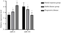
The differences in serum miR-21 and miR-145 expression among the three groups were statistically different (p<0.05). The serum miR-21 levels in patients within the stable disease group and the disease progression group were significantly higher compared to those in the partial response group, while the expression of miR-145 was notably lower when compared to the partial response group (p<0.05). The serum miR-21 levels in patients in the disease progression group were dramatically increased compared to those in the stable disease group, and the expression of miR-145 was significantly reduced in comparison to that of the stable disease group (p<0.05) (Table II and Figure 1).
Analysis of predicting factors for chemotherapy
The age, degree of differentiation (poor differentiation), tumor stage (III-IV), presence of lymphatic metastasis (yes), presence of refractory ascites (yes), miR-21 expression, and miR-145 expression in individuals in the deceased group were statistically different from those in the survival group (p<0.05), while there was no statistically difference in BMI, menopause, and pathological types between the two groups (p>0.05) (Table III).
Multivariate logistic regression analysis
Age, differentiation degree, tumor stage, lymph node metastasis, refractory ascites, miR-21 expression and miR-145 expression were used as independent variables and assigned values for multi-factor logistic regression analysis (Table IV).
Multivariate logistic regression analysis revealed that age, degree of differentiation, tumor stage, lymph node metastasis, refractory ascites, miR-21 expression, and miR-145 expression can be used as independent risk factors affecting the prognosis of patients with ovarian carcinoma undergoing chemotherapy (Table V).
Analysis of diagnostic value of serum miR-21 and miR-145 in the prognostic assessment of patients with ovarian cancer receiving chemotherapy
The ROC analysis showed that the AUC for serum miR-21, miR-145, and their combined assessment in the prognostic evaluation of ovarian cancer patients undergoing chemotherapy were 0.875, 0.890, and 0.898, respectively. The sensitivities for these markers were 86.10%, 86.10%, and 89.90%, respectively, while the specificities were 77.70%, 85.40%, and 89.30%, respectively (Table VI and Figure 2). The analysis indicated that the combined detection of serum miR-21 and miR-145 outperformed the use of a single biomarker in terms of AUC, sensitivity, and specificity.

DISCUSSION
In recent years, numerous approaches have emerged for clinical management of ovarian cancer patients, leading to great improvement in the clinical outcome. However, patients still face a substantial risk of recurrence, and there has been no significant improvement in their overall survival rates (Pergialiotis et al. 2020, Thigpen 2015). Therefore, the exploration of sensitivity indicators tightly linked to the clinical outcomes of ovarian carcinoma patients plays a pivotal role in the development of effective clinical treatment strategies.
Our current study aimed to analyze and explore the predictive value of miR-21 and miR-145 in the efficacy and outcomes of ovarian patients receiving standard chemotherapy. The outcomes of the research revealed that the levels of serum miR-21 were remarkably higher in the stable disease set and the disease progression set compared to the partial response set, while the expression of miR-145 was remarkably lower (p<0.05). The expression of serum miR-21 was also higher in the disease progression set compared to the stable disease set, while the expression of miR-145 was lower (p<0.05). These results indicated that the expression levels of miR-21 and miR-145 were associated with chemotherapy efficacy in patients with ovarian carcinoma. This phenomenon can be attributed to the abnormal increase in the expression of miR-21, which may enhance the proliferative and invasive capabilities of tumor cells. As a result, patients with ovarian cancer may not fully benefit from standard chemotherapy dosages, leading to a decreased effectiveness of the treatment (Rodrigues et al. 2023). miR-145 can remarkably reduce the number of tumor cells and inhibit stemness of cancer stem cells. The tumor suppressor activity declines along with abnormal reduction of miR-145, resulting in enhanced proliferation and differentiation of tumor cells. Additionally, miR-145 also serves as a crucial factor in promoting tumor cell migration and invasion, ultimately leading to suboptimal treatment outcomes for patients. Moreover, miR-145 is vital in enhancing the migration and invasion of tumor cells, which can lead to less effective treatment results for patients (Wang & Zhang 2022). Logistic multifactor analysis revealed that variables such as age, degree of differentiation, tumor stage, lymph node metastasis, refractory ascites, and the expression levels of miR-21 and miR-145 can independently serve as potential risk factors affecting the prognosis of patients with ovarian cancer receiving chemotherapy. An increase in age may lead to reduced resistance to therapies and declined immune function, thereby decreasing a patient’s ability to withstand chemotherapy. This reduction in tolerance can subsequently influence the effectiveness of chemotherapy and negatively affect the overall outcome of the disease for the patient (Cheng et al. 2023, McLean et al. 2010, Van Walree et al. 2019). Poor differentiation, higher tumor stages, and lymphatic metastasis can contribute to the progression of ovarian cancer, adversely affecting ovarian function. These factors present significant challenges to clinical management and result in unfavorable prognostic evaluations for patients (McCluggage et al. 2023, Neal et al. 2007, Stavros et al. 2023). Researchers have reported that high expression of miR-21 promotes physiological processes like tumor cell growth, migration and infiltration by activating the Wnt pathway and upregulating variant CD44 (CD44v6) (Wang et al. 2019, Song et al. 2020). miR-145 have been implicated as a tumor suppressor by targeting and modulating the expression of the sex-determining region Y box protein 11 (SOX11) gene, thereby inhibiting the proliferation of tumor cells (Salas-Huenuleo et al. 2022).
The ROC analysis indicated that serum miR-21, miR-145, and their combined use showed potent diagnostic performance for the prognostic evaluation of ovarian cancer patients undergoing chemotherapy. The improved diagnostic accuracy from their joint detection stemmed from leveraging the strengths of each individual marker. The simultaneous analysis of serum miR-21 and miR-145 offered a more detailed basis for clinically evaluating the prognosis of ovarian cancer patients, thereby enhancing the efficacy of this singular approach. This highlights the significant benefit of utilizing both markers in conjunction (Qiu & Weng 2022, Zhao et al. 2021).
The study on the prognostic significance of serum miR-21 and miR-145 in ovarian cancer patients provides valuable insights but also has some limitations. Firstly, the sample size of 139 patients, while reasonable, may still be considered relatively small for biomarker validation studies, potentially limiting the generalizability of the findings. Another limitation is the study’s focus on only two microRNAs; considering the complex nature of ovarian cancer and its biomarker landscape, the inclusion of additional relevant microRNAs could potentially enhance the predictive accuracy and comprehensiveness of the study. Lastly, the study could benefit from longer follow-up periods to better understand the long-term prognostic implications of these biomarkers in ovarian cancer management.
Reduced expression of serum miR-21 and increased expression of miR-145 are linked to favorable treatment responses and clinical prognoses in patients with ovarian cancer. These biomarkers can act as a supplementary approach for evaluating the effectiveness of treatment and the overall prognostic outlook for individuals with ovarian cancer.
Acknowledgements
This study was supported by Dongguan Science and Technology of Social Development Program (202050715001215).
References
-
ARGHIANI N & MATIN MM. 2021. miR-21: A Key Small Molecule with Great Effects in Combination Cancer Therapy. Nucleic Acid Ther 31: 271-283. https://doi.org/10.1089/nat.2020.0914.
» https://doi.org/10.1089/nat.2020.0914 - BAUTISTA-SÁNCHEZ D ET AL. 2020 The Promising Role of miR-21 as a Cancer Biomarker and Its Importance in RNA-Based Therapeutics. Mol Ther Nucleic Acids 20: 409-420. https://doi.org/10.1016/j.omtn.2020.03.003.
-
CHEN S ET AL. 2022. Mechanisms Controlling MicroRNA Expression in Tumor. Cells 11: 2852. https://doi.org/10.3390/cells11182852.
» https://doi.org/10.3390/cells11182852 -
CHENG H ET AL. 2023. Nomograms for predicting overall survival and cancer-specific survival in elderly patients with epithelial ovarian cancer. J Ovarian Res 16: 75. https://doi.org/10.1186/s13048-023-01144-y.
» https://doi.org/10.1186/s13048-023-01144-y -
ELIES A ET AL. 2018. The role of neoadjuvant chemotherapy in ovarian cancer. Expert Rev Anticancer Ther 18: 555-566. https://doi.org/10.1080/14737140.2018.1458614.
» https://doi.org/10.1080/14737140.2018.1458614 -
FUJIWARA K ET AL. 2021. Olaparib plus bevacizumab as maintenance therapy in patients with newly diagnosed, advanced ovarian cancer: Japan subset from the PAOLA-1/ENGOT-ov25 trial. J Gynecol Oncol 32: e82. https://doi.org/10.3802/jgo.2021.32.e82.
» https://doi.org/10.3802/jgo.2021.32.e82 -
GONZÁLEZ-MARTÍN A ET AL. 2024. Progression-free survival and safety at 3.5 years of follow-up: results from the randomized phase 3 PRIMA/ENGOT-OV26/GOG-3012 trial of niraparib maintenance treatment in patients with newly diagnosed ovarian cancer - a plain language summary. Future Oncol. Advance online publication. https://doi.org/10.2217/fon-2023-0782
» https://doi.org/10.2217/fon-2023-0782 -
HARTER P ET AL. 2022. Efficacy of maintenance olaparib plus bevacizumab according to clinical risk in patients with newly diagnosed, advanced ovarian cancer in the phase III PAOLA-1/ENGOT-ov25 trial. Gynecol Oncol 164: 254-264. https://doi.org/10.1016/j.ygyno.2021.12.016.
» https://doi.org/10.1016/j.ygyno.2021.12.016 - JIANG G, MU J, LIU X, PENG X, ZHONG F, YUAN W, DENG F, PENG X, PENG S & ZENG X. 2020 Prognostic value of miR-21 in gliomas: comprehensive study based on meta-analysis and TCGA dataset validation. Sci Rep 10(1): 4220. https://doi.org/10.1038/s41598-020-61155-3.
-
JIN W, LUO W, FANG W, WANG Y, WANG L, SHEN Q, LIU W & ZHANG H. 2019. miR-145 expression level in tissue predicts prognosis of patients with esophageal squamous cell carcinoma. Pathol Res Pract 215(6): 152401. https://doi.org/10.1016/j.prp.2019.03.029.
» https://doi.org/10.1016/j.prp.2019.03.029 -
LIM MC, CHANG SJ, PARK B, YOO HJ, YOO CW, NAM BH & PARK SY. 2022. Survival After Hyperthermic Intraperitoneal Chemotherapy and Primary or Interval Cytoreductive Surgery in Ovarian Cancer: A Randomized Clinical Trial. JAMA Surg 157: 374-383. https://doi.org/10.1001/jamasurg.2022.0143.
» https://doi.org/10.1001/jamasurg.2022.0143 -
MCCLUGGAGE WG ET AL. 2023. Well-differentiated Sertoli-Leydig Cell Tumors (SLCTs) Are Not Associated With DICER1 Pathogenic Variants and Represent a Different Tumor Type to Moderately and Poorly Differentiated SLCTs. Am J Surg Pathol 47: 490-496. https://doi.org/10.1097/PAS.0000000000002010.
» https://doi.org/10.1097/PAS.0000000000002010 -
MCLEAN KA, SHAH CA, THOMPSON SA, GRAY HJ, SWENSEN RE & GOFF BA. 2010. Ovarian cancer in the elderly: outcomes with neoadjuvant chemotherapy or primary cytoreduction. Gynecol Oncol 118: 43-46. https://doi.org/10.1016/j.ygyno.2010.03.002.
» https://doi.org/10.1016/j.ygyno.2010.03.002 -
MIZUNO T ET AL. 2020. Neoadjuvant chemotherapy promotes the expression of HER3 in patients with ovarian cancer. Oncol Lett 20: 336. https://doi.org/10.3892/ol.2020.12200.
» https://doi.org/10.3892/ol.2020.12200 - NEAL RD, ALLGAR VL, ALI N, LEESE B, HEYWOOD P, PROCTOR G & EVANS J. 2007. Stage, survival and delays in lung, colorectal, prostate and ovarian cancer: comparison between diagnostic routes. Br J Gen Pract 57: 212-219. https://bjgp.org/content/57/536/212.full.pdf.
-
PARK J, LEE JB, LIM MC, KIM BG, KIM JW, KIM S, CHOI CH, KIM HS, PARK SY & LEE JY. 2023. Phase II study of durvalumab and tremelimumab with front-line neoadjuvant chemotherapy in patients with advanced-stage ovarian cancer: primary analysis in the original cohort of KGOG3046/TRU-D. J Immunother Cancer 11: e007444. https://doi.org/10.1136/jitc-2023-007444.
» https://doi.org/10.1136/jitc-2023-007444 -
PERGIALIOTIS V, THOMAKOS N, FROUNTZAS M, HAIDOPOULOS D, LOUTRADIS D & RODOLAKIS A. 2020. Perioperative blood transfusion and ovarian cancer survival rates: A meta-analysis based on univariate, multivariate and propensity score matched data. Eur J Obstet Gynecol Reprod Biol 252: 137-143. https://doi.org/10.1016/j.ejogrb.2020.06.013.
» https://doi.org/10.1016/j.ejogrb.2020.06.013 -
QIU L & WENG G. 2022. The diagnostic value of serum miR-21 in patients with ovarian cancer: a systematic review and meta-analysis. J Ovarian Res 15: 51. https://doi.org/10.1186/s13048-022-00985-3.
» https://doi.org/10.1186/s13048-022-00985-3 -
RALSER DJ, CONDIC M, EGGER E, KOENSGEN D, MUSTEA A & STOPE MB. 2022. Evaluation of the Diagnostic Potential of Circulating MicroRNAs miR-1 and miR-21 in Patients with Ovarian Cancer. Anticancer Res 42(12): 5839-5845. https://doi.org/10.21873/anticanres.16092.
» https://doi.org/10.21873/anticanres.16092 -
RODRIGUES PM ET AL. 2023. miR-21-5p promotes NASH-related hepatocarcinogenesis. Liver Int 43: 2256-2274. https://doi.org/10.1111/liv.15682.
» https://doi.org/10.1111/liv.15682 -
SALAS-HUENULEO E, HERNÁNDEZ A, LOBOS-GONZÁLEZ L, POLAKOVIČOVÁ I, MORALES-ZAVALA F, ARAYA E, CELIS F, ROMERO C & KOGAN MJ. 2022. Peptide targeted gold nanoplatform carrying miR-145 induces antitumoral effects in ovarian cancer cells. Pharmaceutics 14: 958. https://doi.org/10.3390/pharmaceutics14050958.
» https://doi.org/10.3390/pharmaceutics14050958 -
STAVROS S ET AL. 2023. Inguinal lymph node metastasis as sole manifestation of ovarian/fallopian tube cancer: a review of the literature. J Cancer 14: 3176-3181. https://doi.org/10.7150/jca.88863.
» https://doi.org/10.7150/jca.88863 -
SONG KW, ZHANG QG, TAN WB & FANG YN. 2020. Diagnostic significance of serum miR-26b and miR-21 expressions in ovarian cancer and their associations with clinicopathological characteristics and prognosis of patients. Eur Rev Med Pharmacol Sci 24: 1697-1703. https://doi.org/10.26355/eurrev_202002_20344.
» https://doi.org/10.26355/eurrev_202002_20344 -
SYEDA AZ, LANGDEN SSS, MUNKHZUL C, LEE M & SONG SJ. 2020. Regulatory Mechanism of MicroRNA Expression in Cancer. Int J Mol Sci 21(5): 1723. https://doi.org/10.3390/ijms21051723.
» https://doi.org/10.3390/ijms21051723 -
TALAAT A, HELMY MA & SAADAWY SF. 2022. Evaluation of miRNA-21 and CA-125 as a promising diagnostic biomarker in patients with ovarian cancer. Egypt J Med Hum Genet 23(1). https://doi.org/10.1186/s43042-022-00342-5
» https://doi.org/10.1186/s43042-022-00342-5 -
THIGPEN TJ. 2015. Contemporary phase III clinical trial endpoints in advanced ovarian cancer: assessing the pros and cons of objective response rate, progression-free survival, and overall survival. Gynecol Oncol 136: 121-129. https://doi.org/10.1016/j.ygyno.2014.10.010.
» https://doi.org/10.1016/j.ygyno.2014.10.010 -
VAN WALREE IC, VAN SOOLINGEN NJ, HAMAKER ME, SMORENBURG CH, LOUWERS JA & VAN HUIS-TANJA LH. 2019. Treatment decision-making in elderly women with ovarian cancer: an age-based comparison. Int J Gynecol Cancer 29: 158-165. https://doi.org/10.1136/ijgc-2018-000026.
» https://doi.org/10.1136/ijgc-2018-000026 -
WANG M & ZHANG S. 2022. MiR-145 on the proliferation of ovarian cancer cells by regulating the expression of MMP-2/MMP-9. Cell Mol Biol (Noisy-le-Grand) 67(6): 141-148. https://doi.org/10.14715/cmb/2021.67.6.19.
» https://doi.org/10.14715/cmb/2021.67.6.19 -
WANG Y, YANG X, YUAN M, XIAN S, ZHANG L, YANG D & CHENG Y. 2019. Promotion of ovarian cancer cell invasion, migration and colony formation by the miR‑21/Wnt/CD44v6 pathway. Oncol Rep 42(1): 91-102. https://doi.org/10.3892/or.2019.7153.
» https://doi.org/10.3892/or.2019.7153 -
XU W, HUA Y, DENG F, WANG D, WU Y, ZHANG W & TANG J. 2020. MiR-145 in cancer therapy resistance and sensitivity: A comprehensive review. Cancer Sci 111(9): 3122-3131. https://doi.org/10.1111/cas.14517.
» https://doi.org/10.1111/cas.14517 -
YANG J, ZHANG JY, CHEN J, CHEN C, SONG XM, XU Y & LI J. 2014. Prognostic role of microRNA-145 in various human malignant neoplasms: a meta-analysis of 18 related studies. World J Surg Oncol 12: 254. https://doi.org/10.1186/1477-7819-12-254.
» https://doi.org/10.1186/1477-7819-12-254 -
ZHAO S, ZHANG Y, PEI M, WU L & LI J. 2021. miR-145 inhibits mitochondrial function of ovarian cancer by targeting ARL5B. J Ovarian Res 14(1): 8. https://doi.org/10.1186/s13048-020-00762-0.
» https://doi.org/10.1186/s13048-020-00762-0


