
 Extraction of phenolic compounds from Pfaffia glomerata leaves and evaluation of composition, antioxidant and antibacterial properties
Abstract
This work aimed to evaluate the extraction conditions of total phenolic compounds (TPC) from Pfaffia glomerata leaves (PGLs) and characterize the extract obtained in the best conditions. Aqueous extraction was performed in a Shaker, varying the levels of rotation (100, 150, 200 rpm), temperature (30, 45, 60° C) and mass to volume (w v-1) ratio (1:10, 1:20, 1:30 g ml-1). The variables w v-1 ratio and temperature, and their combination, showed a positive effect (p<0.05) in TPC extraction. The time of extraction increased TPC extraction until 30 min, thereafter, the values decreased. The extraction performed at 60o C, 1:30 g ml-1, 125 rpm and 30 min allowed to reach the maximum TPC content (11.94 mg g-1). This extract contains β-ecdysone (4.64 g 100g-1), a chemical marker of P. glomerata, and, phenolic compounds, as gallic acid (28.51 mg 100g-1) and catechin (24.82 mg 100g-1). PGLs extract exhibits antioxidant activity by the in vitro methods evaluated (iron reduction powder and radical scavenging ability). Antibacterial activity was also detected, being found the minimum inhibitory concentration of 20 mg mL-1 for Escherichia coli and Pseudomonas aeruginosa. Therefore, PGL extract had a potential application as natural antioxidant and antimicrobial in food or pharmaceutical products.
Key words
Brazilian ginseng; phenolic acids; flavonoids; β-ecdysone; Escherichia coli
INTRODUCTION
Pfaffia glomerata, traditionally known as Brazilian ginseng, is a medicinal plant, in which the extracts from the roots are of economic interest, due to their promising applications, both in phytomedicine and phytotherapy (Saldanha et al. 2013). When harvesting P. glomerata roots, the leaves are normally discarded, as their compounds are still little known. In this specie Felipe et al. (2019) reported the presence of phenolic compounds and flavonoids in the stems and inflorescences. There is also the presence of saponins, glycosides of high molecular mass, among which β-ecdysone stands out ([2β 3β 5β 22R]-2,3,14,20,22,25-hexahydroxy-colest-7-en -6-one), as it is found only in P. glomerata. This compound is considered the chemical marker of this specie (Vardanega et al. 2016). Studies revealed the presence of β-ecdysone in all the main organs of Brazilian ginseng, such as roots, stems and inflorescences (Serra et al. 2012).
To obtain active compounds from plants, the extraction conditions should be evaluated such as temperature, pH, ratio between the solvent and the plant matrix, and the properties of the plant material, such as composition and particle size (Majinda 2012). The type of solvent used also plays a fundamental role in the extraction processes. Ethanol and water are the most recommended solvents aiming to carry out sustainable processes, as they are highly efficient and non-toxic (Prat et al. 2016).
Most studies involving P. glomerata focus on extraction conditions to obtain saponins or β-ecdysone (Da Silva et al. 2021, Debien et al. 2015, Martins et al. 2020), but such investigation focused on phenol’s extraction is scarce in literature. Phenolic compounds, widely distributed in plants, can perform a series of biological activities such as antioxidants and antimicrobials, according to their chemical structure (Lobiuc et al. 2023).
Antioxidant action is of great importance, both in food and in the human body, as through the donation of electrons or the sequestration of oxygen, the oxidative effects caused by free radicals are minimized. Antimicrobial activity is related to inhibiting the development of pathogenic and spoilage microorganisms, which can cause foodborne diseases, thus contributing to quality and safety for consumers. Furthermore, due to the resistance to antibacterials and the possibility of toxicity of synthetic additives, the search for natural compounds as food preservatives and antioxidants is increasingly of interest (Takó et al. 2020).
Several studies have reported the antioxidant and antimicrobial activity of other ginseng species, such as Panax ginseng (Lee et al. 2012, 2016, Lim et al. 2009, Pina-Perez et al. 2018, Xue et al. 2017). However, little information is known to date for P. glomerata, especially for the leaves, which are generally burned when harvesting the roots, posing environmental risks (Da Silva et al. 2021, Felipe et al. 2019). Furthermore, unconventional extraction processes, such as high pressure, are not considered necessary to obtain active compounds from P. glomerata (Vardanega et al. 2016); thus, simple, and low-cost processes, such as orbital agitation, could be evaluated.
Therefore, the aim of this work was to study the effect of extraction conditions (rotation, temperature and weight/volume ratio) by orbital shaking on obtaining phenolic compounds from P. glomerata leaves, and, in the best extraction condition, evaluate the phenolic composition, the presence of β-ecdysone, and the antioxidant and antibacterial properties.
MATERIALS AND METHODS
Obtaining and preparing P. glomerata leaves
P. glomerata leaves (PGL) were obtained from producers in the city of Iporã/PR (24°00’11”S 53°42’15”W), where they were harvested manually and washed with potable water. The leaves were dried in an oven with forced air circulation (Marconi, MA35) at 60° C, for 18 h, until reaching a moisture of 10±1% (m m-1). The dried leaves were crushed in an analytical mill (Ika, A11) and the particle size was defined using vibrating sieves, being the material retained in the sieve of 0.15 mm used in the extraction experiments.
Extractions
The extractions were carried out in an orbital shaker (Marconi, MA 42), using water as solvent. A factorial experimental design (Box-Behnken) was followed during extraction, testing three levels of each variable: rotation – X1 (100, 150, 200 rpm), temperature – X2 (30, 45, 60°C) and weight to volume ratio (w v-1) – X3 (1:10; 1:20; 1:30 mg ml-1), with three repetitions at the central point. After each extraction, filtration and analysis of the content of total phenolic compounds (TPC) were carried out.
The TPC content was measured using the Folin-Ciocalteau reagent (10%, v v-1) and sodium carbonate (7.5% w v-1). The absorbance was read in spectrophotometer (Shimadzu, 1900 UV/VIS) at 760 nm (Shahidi & Zhong 2015). Gallic acid solutions (25 to 500 mg L-1) were used to obtain the analytical curve, and the results were expressed as gallic acid equivalent (GAE).
The statistical analysis of the experimental design allows obtaining the condition of maximum TPC extraction, within the tested ranges. In this condition, the extraction was repeated in triplicate, in order to establish whether the data predicted by the mathematical model generated in the analysis corresponds to the experimental data. Under the same condition, tests were carried out to define the effect of extraction time (0, 10, 20, 30, 40, 50 and 60 min), and TPC contents were evaluated in the extracts.
Characterization of the extract obtained under the best experimental condition
The extract obtained under the best extraction conditions, defined both by experimental planning and by studying the effect of time, was analyzed for composition and antioxidant and antibacterial properties, as described below.
Composition
Total flavonoids (TF) content was measured according to Boateng et al. (2008), by the reaction of extract components with NaNO2 (50 g L-1), AlCl3 (100 g L-1) and NaOH (1 M). The absorbance reading was performed at 510 nm and the TF content was calculated based on the quercetin analytical curve and expressed as quercetin equivalent (EQ).
The PGL extract was filtered (PVDF hydrophobic membrane, pore size 0.45μm and 25mm diameter) and analyzed by high-performance liquid chromatography (HPLC) for detection of phenolic compounds and β-ecdysone. The phenolic profile was investigated in the HPLC 20A (Shimadzu), C-18 column (Shim-pack CLC-ODS (H)™, 25 cm x 4.6 mm x 5 mm, Shimadzu), according to Donadone et al. (2020). The mobile phases, ultrapure water and methanol, both acidified with 0.05% and 0.10% (v v-1) formic acid, respectively, were used in gradient elution mode, at 0.8 mL min-1 at 25°C. Detection occurred at wavelengths of 280 and 320 nm (SPD-20A, Shimadzu). For quantification, it was used analytical curves (R2>0.99) with concentrations between 1 to 10 mg mL-1 of the analytical standards: gallic, coumaric, ferulic, caffeic, trans cinnamic acids, and the flavonoids quercetin, catechin and kaempferol, all from Sigma–Aldrich. The recovery of the standards added to the PGL extract was, in media, 94%, and the relative standard deviation was 6%.
The β-ecdysone content was determined according to the method validated by Serra et al. (2012), with few adaptations described by Martins et al. (2020). It was used a HPLC (Jasco LC-4000), with a C18 reverse-phase column (Fortis, 250 × 4.6 nm, 5 μm), which was maintained at 30 °C. The chromatographic run was performed in gradient mode, using methanol (phase A) and ultrapure water (phase B), in gradient mode as follows: 0–5 min, 10–70% A; 5–12 min, 70% A; and 12–15 min, 70–100% A. The flow rate was 1 mL min-1, the injection volume was 20 μL and the compound of interest was read at 245 nm (SPD-10A). The calibration curve was established by the external standard method, using the β-ecdysone (20-hydroxyecdysone, Sigma–Aldrich) with concentrations of 30 to 250 mg L-1, which showed linearity, with slope (a) of and intercept (b) values of 479 and 5157, respectively, and correlation coefficient of 0.99. The recovery test, based on the addition of solutions of β-ecdysone standard (50 and 100 mg L-1) in the extract, showed a median value of 97%, with relative standard deviation of 5%.
Antioxidant activity
The antioxidant properties were measured according Donadone et al. (2020). In the DPPH (2,2-diphenyl-1-picrylhydrazyl) method a stock solution of DPPH (0.4 g L-1) in ethanol was prepared and diluted to obtain an absorbance of 0.80±0.02 at 517 nm. This solution (3.9 mL) was mixed with 0.1 mL of the extract, and after 30 min in the dark, the absorbance was read again. The iron reducing antioxidant power (FRAP) method was used by mixing 2.7 mL of FRAP reagent (acetate buffer, 0.3 mM pH 3.6, TPTZ solution (2,4,6-tri(2-pyridyl)-1,3,5-triazine) 10 mM and 20 mM ferric chloride), 90 µL of the extract and 0.27 mL of distilled water. After homogenization, the sample was incubated at 37 °C for 30 min and reading was performed at 595 nm. The ABTS (2,2’-azino-bis(3-ethylbenzothiazoline-6-sulfonic acid) free radical method was carried out by dissolving 7 mM ABTS in ethanol and 140 mM potassium persulfate until the absorbance reach 0.70±0.05 nm at 734 nm. This solution (3 mL) was mixed with 30 μL of the extract and after 6 min of reaction, the reading was performed again.
The results of the antioxidant activity methodologies were expressed in Trolox equivalent (TE). To this end, analytical curves were prepared containing Trolox (6-hydroxy-2,5,7,8-tetramethylchroman-2-carboxylic acid) in concentrations of 0.10 to 0.80 mM (DPPH), 0.15 to 0.75 mM (FRAP) and 0.10 to 2.0 mM (ABTS).
Antibacterial activity
Antibacterial activity was carried out by the broth microdilution method (CLSI 2015) modified for natural products. Screening was carried out to determine the minimum inhibitory concentration (MIC) of each microorganism through serial microdilution, in 96-well microplates. For each microorganism, a standardized suspension was obtained, according to the 0.5 McFarland scale (1.5 x 10⁸ UFC mL-1) in 0.85% (w v-1) saline solution. The inoculum concentration was adjusted with Muller Hinton broth to 1.5 x 10⁷ UFC mL-1.
P. glomerata extract was dissolved in sterile distilled water and evaluated in the range of 80 to 0.3125 mg mL-1. Sodium nitrite is commonly used as a food additive, so it was used as commercial standard, for comparison. This additive was dissolved in sterile distilled water and was evaluated in the range of 100 to 0.19 mg mL-1. All samples were evaluated in 50 μL of solution (culture medium and samples). After serial dilution, 50 µL of the inoculum was added to each well, reaching a concentration of 1.5 x 10⁵ UFC mL-1, and subjected to incubation at 35 °C for 24 h. Reading was performed by adding 20 μL of 1% (w v-1) 2,3,5-triphenyltetrazolium chloride developer to each well followed by incubating the microplates at 35° C for 20 min. The MIC was defined as the lowest concentration that resulted in growth inhibition by visual inspection. The minimum bactericidal concentration (MBC) was determined by subculturing 5 µL of each well on Muller Hinton agar, subsequently defined as the lowest concentration that kills ≥ 99.9% of bacteria after 24 h of incubation at 35 °C.
Statistical data analysis
All experiments were carried out in triplicate, and the results were expressed as mean ± standard deviation, on a dry basis (b.s.). The analysis of data obtained in the Box-Behnken experimental design, using a significance level of 5% (p=0.05), was carried out using the software Statistica 7.0 (StatSoft TM, Inc). In the same software, the results obtained for the extraction characterization were compared using analysis of variance (ANOVA) followed by the Tukey test (p=0.05).
RESULTS AND DISCUSSION
Effect of extraction conditions on TPC contents
The results regarding the TPC content extracted from PGL, following the Box-Behnken experimental design, are expressed in Table I. The TPC contents varied from 0.87 to 11.63 mg g-1, which demonstrates that the variables influenced in the extraction of these compounds. Furthermore, it is important to highlight tests 13, 14 and 15 (central points), showed low variation between them, indicating a good repeatability of the experiment. Malathy et al. (2020) carried out aqueous extraction of another ginseng specie (Panax ginseng) and detected in the leaves 12.66 mg g-1 of TPC, a similar result to that obtained in the present study. According to these authors, the TPC content in the leaves was higher than that found in the roots (8.21 mg g-1), showing the importance of investigating unconventional parts of the plants. This finding can be reinforced by observing other studies using P. ginseng roots, with maximum TPC levels, using water as solvent, were 6.6 mg g-1 (Kim et al. 2007) and 2.0 mg g-1 (Zhang et al. 2018), even when using other extraction technologies, ultrasound and subcritical water, respectively. Such values are lower than the results obtained in this study, especially in the condition of higher TPC extraction (11.63 mg g-1).
Experimental conditions of the Box-Behnken experimental design and the results for total phenolic content (TPC) extracted from P. glomerata leaves.
Table II shows the influence of each variable (X1, X2 and X3) in the TPC extraction from PGL. It was observed positive effects (p<0.05) for the variables studied, being quadratic for rotation (X1), linear for temperature (X2), and both quadratic and linear for w v-1 ratio (X3). These results demonstrated that increasing the level of the variables, there is an increase in TPC extraction, however, for rotation and the w v-1 ratio this effect occurs until a certain level, after which there is a tendency of TPC reduction (quadratic effect).
Estimated effects for the independent variables rotation (X1), temperature (X2) and weight to volume ratio (X3) in the extraction of total phenolic content extracted from P. glomerata leaves.
The combination of temperature and w v-1 ratio also promoted a positive effect (p<0.05) on TPC extraction. However, the combination of the w v-1 ratio and rotation showed a negative effect (p<0.05), indicating that the combination of these variables at their highest levels reduces TPC extraction. Similarly, Saravana et al. (2016) reported positive influence of the w v-1 ratio and temperature in the TPC extraction from ginseng by-products using pressurized hot water.
Lee et al. (2016) reported that temperature had a positive linear effect increasing the TPC content of Korean red ginseng, for example, there was a 53% increase in CFT content when increasing the extraction temperature from 30 to 90 °C. In the present work, the increase in TPC content was 30% and 50%, when comparing experiments 1 and 3, and 2 and 4, respectively, in which the extraction temperature was from 30 to 60 °C, with the other extraction conditions remaining constant. Extraction conducted at a higher temperature can reduce the viscosity and surface tension of the solvent, which allows faster TPC removal, or it can partially hydrolyze some lignin fragments, increasing TPC levels (Beaufils et al. 2021)
The effects of temperature and rotation may be related to the fact that their increase promotes the diffusion and solubility of phenolic compounds, thus facilitating the penetration of the solvent, making extraction more efficient. However, extraction is facilitated by the agitation process until the moment that adsorption decreases, due to the number of active sites becoming constant and maximum elimination of compounds is then achieved, and this fact could explain the quadratic effect of rotation obtained in this study (Chen et al. 2014).
The effect of the w v-1 ratio is normally associated with the fact that the higher volume of solvent used in the extraction can promote an increase/swelling of the solid matrix, causing a rupture of the walls, allowing greater release of bioactive compounds into the aqueous medium (Jin et al. 2011). In this study, the increase of the w v-1 ratio from 1:10 to 1:30 (remaining the other extraction conditions constants) increased the TPC contents from 2.2 times (experiments 6 and 8) to 5.5 times (experiments 10 and 11).
Based on Table II, taking into account the linear and quadratic regression coefficients, considering only the parameters with significant effect, it was possible to obtain the second-order mathematical model equation for the TPC content (Eq. 1).
Where X1 is the rotation (rpm), X2 temperature (°C), X3 w v-1 ratio (g ml-1).
The model (Eq. 1) was considered valid because the ANOVA data showed a higher F value (55.86) than the critical F value (3.58). The coefficient of determination (R2) was 0.96. The statistical analysis and the F-test showed that the model adequately represented the experimental data in the range tested, allowing the maximum TPC values to be determined.
According to the result predicted by the mathematical model (Eq. 1), the maximum TPC content were obtained using the highest levels (1.0) of the variables temperature (60 °C) and w/v ratio (1:30 g ml-1), while rotation must be applied at low level (-0.5 or 125 rpm). Thus, the value predicted by the mathematical model was 11.29 mg g-1. This value did not differ (p<0.05) from the value obtained experimentally, 11.94 ± 0.09 mg g-1, showing that the model was well adjusted to represent the experimental data.
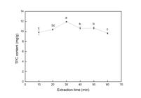
In view of this, the effect of extraction time was evaluated, fixing the other parameters (60° C, 1:30 g mL-1, 125 rpm) as can be seen in Figure 1. It was noted that time had a quadratic effect, so that the TPC content increased during the 10 to 30 min extraction interval. After this period, the TPC content decreased, remaining constant between 40 and 50 min, and decreasing again after 60 min of extraction. This suggests that prolonged exposure to treatments involving high temperatures can promote the degradation of phenolic compounds. Lee et al. (2016) reported that 30 min was sufficient for extracting TPC from Korean red ginseng.
Effect of time extraction on the total phenolic compounds content in P. glomerata leaves in the conditions defined by Box-Behnken experimental design (125 rpm, 60 °C, 1:30 g mL-1).
Composition of PGL extract
The extract of P. glomerata leaves, obtained using the best extraction conditions (125 rpm, 60 °C, 1:30 g mL-1 and 30 min) presented flavonoids, phenolic acids and β-ecdysone.
According to Lee et al. (2012), the extract obtained from the leaves of the P. ginseng showed a total flavonoid content corresponding to ~20% of the TPC content, similar to the value found in the present study (18.34%). Among the flavonoids identified in PGL, catechin was the predominant one, followed by kaempferol and quercetin. Catechin is an important member of the polyphenols in green teas, and it has strong antioxidant potential (Bai et al. 2021).
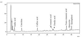
Figure 2 shows the chromatogram of PGL extract. It is composed by gallic, trans-cinamic, ferrulic, coumaric and cafeic acids, whose contents are higher than those obtained for flavonoids. Chung et al. (2016) found that P. ginseng leaves had more phenolic acids, but lower flavonoids content than the roots. Gallic acid, the main phenolic acid found in PGL extract, is a polyphenol, with three hydroxyl groups bound to the aromatic ring, known to inhibit oxidative stress and to have pharmacological activities, such as antioxidant, anti-inflammatory and anticancer (Bai et al. 2021).
The β-ecdysone content in PGL extract (4.64 g 100g-1) was similar than that found in the roots (5.0 g 100g-1), according to Debien et al. (2015). β-ecdysone can exhibit anti-diabetic, immunomodulatory, hepatoprotective, antiarrhythmic and cholesterol-lowering properties (Da Silva et al. 2021). The synthesis of this compound is very difficult, so P. glomerata is cultivated mainly for β-ecdysone extraction from its roots. In the harvest of the roots the aerial parts are usually burned, causing an environmental problem, that could be avoided using them for active compounds extraction (Da Silva et al. 2021, Felipe et al. 2019).
Antioxidant activity of PGL extract
The antioxidant activity of PGL extract is shown in Table IV. Phenolic hydroxyl compounds, as acid phenols and flavonoids, possess electron and/or hydrogen donor ability and hence, exhibit antioxidant activity (Bai et al. 2021). The PGL extract showed antioxidant activity in all the methods evaluated; the highest antioxidant activity was observed for FRAP, followed by DPPH, and after by ABTS. Malathy et al. (2020) reported that leaves of P. ginseng showed higher antioxidant activity by nitric oxide free radical method, than the roots.
Another factor that may influence the antioxidant activity results is the solvent used in the extraction. Kim et al. (2007) reported that the antioxidant activity by the DPPH method was 1.5 to 5 times higher when only water was used to extract the P.ginseng roots, in relation to mixtures of water and ethanol. It was not found studies about antioxidant activity for aqueous extracts from P. glomerata leaves, so, this work contributes to the knowledge of the properties of this kind of extract.
Antibacterial activity of PGL extract
Regarding the antibacterial activity of PGL extract, the MIC values ranged from 20.00 to 80.00 mg mL-1, while for the commercial standard, sodium nitrite, the values were within 12.50 and 100.00 mg mL-1 (Table V).
Minimum inhibitory concentration (MIC) of the extract from P.glomerata leaves (PGL) and commercial standard sodium nitrite.
For E. coli and S. typhi PGL extract showed MIC of 20.00 and 80.00 mg mL-1, respectively, these values were lower than those found for sodium nitrite (25 and 100 mg mL-1, respectively). For P. aeruginosa, PGL showed a MIC (20 mg mL-1) 1.6 times higher than the standard (12.50 mg mL-1). In relation to S. aureus and B. cereus PGL extract exhibit MIC values 1.2 and 6.4 times higher than the MIC values of sodium nitrite.
Sodium nitrite is an additive that has antimicrobial and antioxidant action in foods, but depending on the concentration, it can cause toxic effects in the human body such as methemoglobinemia and teratogenic effects (Hamdi et al. 2018). Therefore, the use of natural compounds to replace synthetic additives such as sodium nitrite is of interest to the food industry. In relation to MBC, the values were higher than 80.00 mg mL-1 (data not showed) for all species tested, showing that at the concentrations evaluated, the extract had a bacteriostatic action.
To date, no reports of antimicrobial activity have been found for P. glomerata, but there are researches about other species. The glycolic extract of P. paniculata showed antibacterial activity against both S. aureus and S. epidermidis, with a MIC of 25 mg mL-1 for both (Miranda et al. 2024), value similar to the one found in the present study for P. aeruginosa and E. coli. ginseng (Panax quinquefolius L.) extract showed growth inhibitory potential only against vegetative Gram (-) bacteria, being the concentration of 10% (w v-1) able to inhibit E. coli growth (105 - 107 UFC mL-1) (Pina-Perez et al. 2018). Xue et al. (2017) obtained heat-transformed saponins from Panax quinquefolius leaf-stem extract, and reported antimicrobial activity in relation to F. nucleatum ,C. perfringens, and P. gingivalis. Extracts of P. ginseng roots showed antimicrobial activity against P. aeruginosa ,S. typhimurium ,S. aureus ,B. cereus, and E. coli (Lim et al. 2009). While extracts of stems-leaves, obtained by subcritical water extraction, inhibited the growth of B. cereus ,S. enteritidis ,E. coli and Listeria monocytogenes (Lee et al. 2012)
Although this study only tested the crude extract of PGL, some components detected (Table III) may possibly be correlated with antibacterial effects. As example, catechin can inhibit some Gram-positive and Gram-negative bacteria due to its ability to adsorb onto the membrane surface, deteriorating it and causing its rupture (Ansari et al. 2017). Gallic acid may also be associated with antibacterial action, as it shows activity against E. coli and S. aureus with MICs of 0.25 and 0.50 mg mL-1, respectively (Carvalho et al. 2018). Phenolic acids can cause a decrease in extracellular pH, alter membrane potential and cause leakage of cellular constituents, as well as denaturing proteins in the cytoplasm (Lobiuc et al. 2023). According to Pina-Perez et al. (2018) ginsenosides, such as β-ecdysone, have been reported to be the major antimicrobial compounds in ginseng.
Further studies should be carried out to confirm the main components responsible for the antibacterial effect of the PGL extract obtained in this study, as well as to fractionate or purify the extract in order to maximize this activity. In any case, the results are promising, since the PGL extract is easy and inexpensive to obtain and does not require the use of toxic solvents. In addition, the extract is obtained from a natural source, unlike sodium nitrite, which is known to have potential adverse effects on the human body, being linked to diseases such as cancer, allergies and gastrointestinal problems (Hamdi et al. 2018).
CONCLUSIONS
In this study, aqueous extraction by orbital shaking at 125 rpm, 60°C, for 30 min, using w v-1 ratio of 1:30 g mL-1 enabled to reach the highest CFT content (11.62 mg g-1) from PGL, among the conditions evaluated. This extract showed phenolic acids, flavonoids and β-ecdysone in its composition, as well as antioxidant and antibacterial activity. Thus, P. glomerata leaves have the potential to be used as a source of active compounds, that can be obtained by an environmentally and economically extraction method, as presented in this work. The compounds extraction represents an alternative for the integral use of the plant, which can enable the valorization of production chain, avoiding environmental problems caused by burning the aerial parts of P. glomerata.
ACKNOWLEDGMENTS
Authors acknowledge to Coordenação de Aperfeiçoamento de Pessoal de Nível Superior – Brasil (CAPES), finance code 001, and to Conselho Nacional de Desenvolvimento Científico e Tecnológico (CNPq), process 310472/2022-9.
REFERENCES
-
ANSARI F, POURJAFAR H & ESMAILPOUR S. 2017. Study on Citric Acid Production and Antibacterial Activity of Kombucha Green Tea Beverage during Production and Storage. Annu Res & Rev Biol 16: 1-8. http://dx.doi.org/10.9734/ARRB/2017/35664.
» https://doi.org/10.9734/ARRB/2017/35664 -
BAI J, ZHANG Y, TANG C, HOU Y, AI X, CHEN X, ZHANG Y, WANG X & MENG X. 2021. Gallic acid: Pharmacological activities and molecular mechanisms involved in inflammation-related diseases. Biomed Pharmacother 133: 1-14. https://doi.org/10.1016/j.biopha.2020.110985.
» https://doi.org/10.1016/j.biopha.2020.110985 -
BEAUFILS N, BOUCHER J, PEYDECASTAING J, RIGAL L, VILAREM G, VILLETTE MJ, CANDY L & PONTALIER PY. 2021. The effect of time and temperature on the extraction of xylose and total phenolic compounds with pressurized hot water from hardwood species used for pulp and paper production in the South of France. Bioresour Technol Rep 16: e100832. https://doi.org/10.1016/j.biteb.2021.100832.
» https://doi.org/10.1016/j.biteb.2021.100832 -
BOATENG MJ, VERGHESE LT & OGUTU SW. 2008. Effect of processing on antioxidant contents in selected dry beans (Phaseolus spp. L.). LWT - Food Sci Technol 41: 1541-1547. https://doi.org/10.1016/j.lwt.2007.11.025.
» https://doi.org/10.1016/j.lwt.2007.11.025 -
CARVALHO RS, CAROLLO CA, DE MAGALHÃES JC, PALUMBO JMC, BOARETTO AG, NUNES E SÁ IC, FERRAZ AC, LIMA WG, DE SIQUEIRA JM & FERREIRA JMS. 2018. Antibacterial and antifungal activities of phenolic compound-enriched ethyl acetate fraction from Cochlospermum regium (mart. Et. Schr.) Pilger roots: Mechanisms of action and synergism with tannin and gallic acid. S Afr J Bot 114: 181-187. https://doi.org/10.1016/j.sajb.2017.11.010.
» https://doi.org/10.1016/j.sajb.2017.11.010 -
CHEN W, HUANG Y, QI J, TANG M, ZHENG Y, ZHAO S & CHEN L. 2014. Optimization of ultrasound-assisted extraction of phenolic compounds from Areca husk. J Food Process Eng 38: 90-96. https://doi.org/10.1111/j.1745-4549.2012. 00748.x.
» https://doi.org/10.1111/j.1745-4549.2012 -
CHUNG IM, LIM JJ, AHN MS, JEONG HN, AN TJ & KIM SH. 2016. Comparative phenolic compound profiles and antioxidative activity of the fruit, leaves, and roots of Korean ginseng (Panax ginseng Meyer) according to cultivation years. J Ginseng Res 40: 68-75. https://doi.org/10.1016/j.jgr.2015.05.006.
» https://doi.org/10.1016/j.jgr.2015.05.006 - CLSI - CLINICAL AND LABORATORY STANDARDS INSTITUTE. 2015. Methods for dilution antimicrobial susceptibility tests for bacteria that grow aerobically; Approved Standard, CLSI document M07-A10. Wayne, USA.
-
DA SILVA TFO, YAMAGUCHI CS, RIBEIRO STC, DA SILVA AVINCOLA A, PILAU EJ, PORTO C, DE OLIVEIRA AJ & GONÇALVES RAC. 2021. Adventitious root culture of Pfaffia glomerata (Spreng.) Pedersen in a roller bottle system: An alternative source of β-ecdysone. Phytochem Lett 43: 1-7. https://doi.org/10.1016/j.phytol.2021.02.009.
» https://doi.org/10.1016/j.phytol.2021.02.009 -
DEBIEN ICN, VARDANEGA R, SANTOS DT & MEIRELES MAA. 2015. Pressurized liquid extraction as a promising and economically feasible technique for obtaining Beta-ecdysone- rich extracts from Brazilian ginseng (Pfaffia glomerata) Roots. Sep Sci Technol 50: 11. https://doi.org/10.1080/01496395.2014.975361.
» https://doi.org/10.1080/01496395.2014.975361 -
DONADONE DBS, GIOMBELLI C, SILVA DLG, STEVANATO N, SILVA C & BARROS BCB. 2020. Ultrasound-assisted extraction of phenolic compounds andsoluble sugars from the stem portion of peach palm. J Food Process Pres 44: e14636. https://doi.org/10.1111/jfpp.14636.
» https://doi.org/10.1111/jfpp.14636 -
FELIPE SHS ET AL. 2019. Salinity-induced modifications on growth, physiology and 20-hydroxyecdysone levels in Brazilian-ginseng [Pfaffia glomerata (Spreng.) Pedersen]. J Plant Biochem Physiol 140: 43-54. https://doi.org/10.1016/j.plaphy.2019.05.002.
» https://doi.org/10.1016/j.plaphy.2019.05.002 -
HAMDI M, NASRI R, DRIDI N, HAFEDH M, ASHOUR L & NASRI M. 2018. Improvement of the quality and the shelf life of reduced-nitrites turkey meat sausages incorporated with carotenoproteins from blue crabs shells. Food Control 91: 148-159. https://doi.org/10.1016/j.foodcont.2018.03.048.
» https://doi.org/10.1016/j.foodcont.2018.03.048 - JIN R, FAN L & AN X. 2011. Pré-tratamento líquido iônico assistido por microondas de plantas medicinais para extração rápida de solventes de ingredientes ativos. Purif. Tecnol 83: 45-49.
-
KIM S, MURTHY HN, HAHN E, LEE HL & PAEK K. 2007. Parameters affecting the extraction of ginsenosides from the adventitious roots of ginseng (Panax ginseng C.A. Meyer). Sep Purific Technol 56: 401-406. https://doi.org/10.1016/j.seppur.2007.06. 014.
» https://doi.org/10.1016/j.seppur.2007.06 -
LEE KA, KIM WJ, KIM HJ, KIM K & PAIK H. 2012. Antibacterial activity of ginseng (Panax ginseng C. A. Meyer) stems–leaves extract produced by subcritical water extraction. Int J Food Sci Technol 48: 947-953. http://dx.doi.org/10.1111/ijfs.12046.
» https://doi.org/10.1111/ijfs.12046 -
LEE LW, MO EJ, CHOI EJ, JO JH, JANG H, JEONG JY, JIN Q, CHUNG HN, HWANG BY & LEE MK. 2016. Effect of Korean Red ginseng extraction conditions on antioxidant activity, extraction yield, and ginsenoside Rg1 and phenolic content: optimization using response surface methodology. J Ginseng Res 40: 229-236. https://doi.org/10.1016/j.jgr.2015.08.001.
» https://doi.org/10.1016/j.jgr.2015.08.001 - LIM JK, KANG HJ, KANG SN & LEE BY. 2009. Antioxidant and antimicrobial activities of various solvent fractions of fine ginseng root. Food Sci Biotechnol 18: 513-518.
-
LOBIUC A, PAVĂL NE, MANGALAGIU II, GHEORGHIȚĂ R, TELIBAN GC, AMĂRIUCĂI-MANTU D & STOLERU V. 2023. Future Antimicrobials: Natural and Functionalized Phenolics. Molecules 28: e1114. https://doi.org/10.3390/mole cules28031114.
» https://doi.org/10.3390/mole cules28031114 -
MAJINDA RRT. 2012. Extraction and isolation of saponins. In: Sarker SD & Nahar L (Eds), Methods Mol Biol 864: 415-426. https://doi.org/10.1007/978-1-61779-624-1_16.
» https://doi.org/10.1007/978-1-61779-624-1_16 -
MALATHY R, PRABAKARAN M, KALAISELVI K, CHUNG IM & KIM SH. 2020. Comparative Polyphenol Composition, Antioxidant and Anticorrosion Properties in Various Parts of Panax ginseng Extracted in Different Solvents. Appl Sci 11: 93. http://dx.doi.org/10.3390/app11010093.
» https://doi.org/10.3390/app11010093 -
MARTINS N, ANDRICH F, MARTINS D, CALAÇA G & SAKAI O. 2020. Determination of β-ecdysone in infusions of different organs of Brazilian ginseng (Pfaffia glomerata) by high-performance liquid chromatography. Rev Mundi Eng Tecnol Gest 5: 1-14. https://doi.org/10.33448/rsd-v10i4.14147.
» https://doi.org/10.33448/rsd-v10i4.14147 -
MIRANDA DG, CARROUEL F, SILVA TCA, ROZZATO MC, HASNA AA, SANTOS CER, MORAIS FV, OLIVEIRA LD & RAMOS LP. 2024. New Insights into Cutaneous Asepsis: Synergism between Pfaffia and Rosemary Extracts. Antibiotics 13: 226. https://doi.org/10.3390/antibiotics13030226.
» https://doi.org/10.3390/antibiotics13030226 -
PINA-PÉREZ MC, RIVAS A, MARTÍNEZ A & RODRIGO D. 2018. Effect of thermal treatment, microwave, and pulsed electric field processing on the antimicrobial potential of açaí (Euterpe oleracea), stevia (Stevia rebaudiana Bertoni), and ginseng (Panax quinquefolius L.) extracts. Food Control 90: 98-104. https://doi.org/10.1016/j.foodcont.2018.02.022.
» https://doi.org/10.1016/j.foodcont.2018.02.022 - PRAT D, WELLS A, HAYLER J, SNEDDON H, MCELROY CR, ABOU-SHEHADA S & DUNN PJ. 2016. CHEM21 selection guide of classical- and less classical-solvents. Green Chem 18: 288-296. http://xlink.rsc.org/?DOI=c5gc01008j.
-
SALDANHA CW, OTONI CG, NOTINI MM, KUKI KN, CRUZ ACF, NETO AR, DIAS LLC & OTONI WC. 2013. A CO2-enriched atmosphere improves in vitro growth of Brazilian ginseng (Pfaffia glomerata (Spreng.) Pedersen). In Vitro Cell Deve Biol Plant 49: 433-444. https://doi.org/10.1007/s11627-013-9529-5.
» https://doi.org/10.1007/s11627-013-9529-5 -
SARAVANA PS, GETACHEW AT, AHMED T, CHO Y, LEE Y B & CHUN BS. 2016. Optimization of phytochemicals production from the ginseng by-products using pressurized hot water: Experimental and dynamic modelling. J Eng Biochem 113: 141-151. https://doi.org/10.1016/j.bej.2016.06.006.
» https://doi.org/10.1016/j.bej.2016.06.006 -
SERRA LZ, FELIPE DF & CORTEZ DAG. 2012. Quantificação de ÿ-ecdisona em diferentes partes de Pfaffia glomerata por HPLC. Rev Bras Farmacogn 22: 1319-1354. https://doi.org/10.1590/S0102-695X2012005000114.
» https://doi.org/10.1590/S0102-695X2012005000114 -
SHAHIDI F & ZHONG Y. 2015. Measurement of antioxidant activity. J Funct Foods 18: 757-781. https://doi.org/10.1016/j.jff.2015.01.047.
» https://doi.org/10.1016/j.jff.2015.01.047 -
TAKÓ M, KEREKES EB, ZAMBRANO C, KOTOGÁN A, PAPP T, KRISCH J & VÁGVÖLGYI C. 2020. Plant phenolics and phenolic-enriched extracts as antimicrobial agents against food-contaminating microorganisms. Antioxidants 9: 165. https://doi.org/10.3390%2Fantiox9020165.
» https://doi.org/10.3390%2Fantiox9020165 -
VARDANEGA R, CARVALHO PIN, ALBARELLI JQ, SANTOS DT & MEIRELES MAA. 2016. Techno-economic evaluation of obtaining Brazilian ginseng extracts in potential production scenarios. Food Bioprod Process 101: 45-55. https://doi.org/10.1016/j.fbp.2016.10.010.
» https://doi.org/10.1016/j.fbp.2016.10.010 -
XUE P, YAO Y, YANG X, FENG J & REN G. 2017. Improved antimicrobial effect of ginseng extract by heat transformation. J Ginseng Res 41: 80-187. https://doi.org/10.1016/j.jgr.2016.03.002.
» https://doi.org/10.1016/j.jgr.2016.03.002 -
ZHANG Y, TAHA AA, YIN Y, LI X, CHEN X & MA C. 2018. Subcritical water extraction of bioactive components from ginseng roots (Panax ginseng C.A. Mey). Ind Crops Prod 117: 118-127. https://doi.org/10.1016/j.indcrop.2018.02.079.
» https://doi.org/10.1016/j.indcrop.2018.02.079

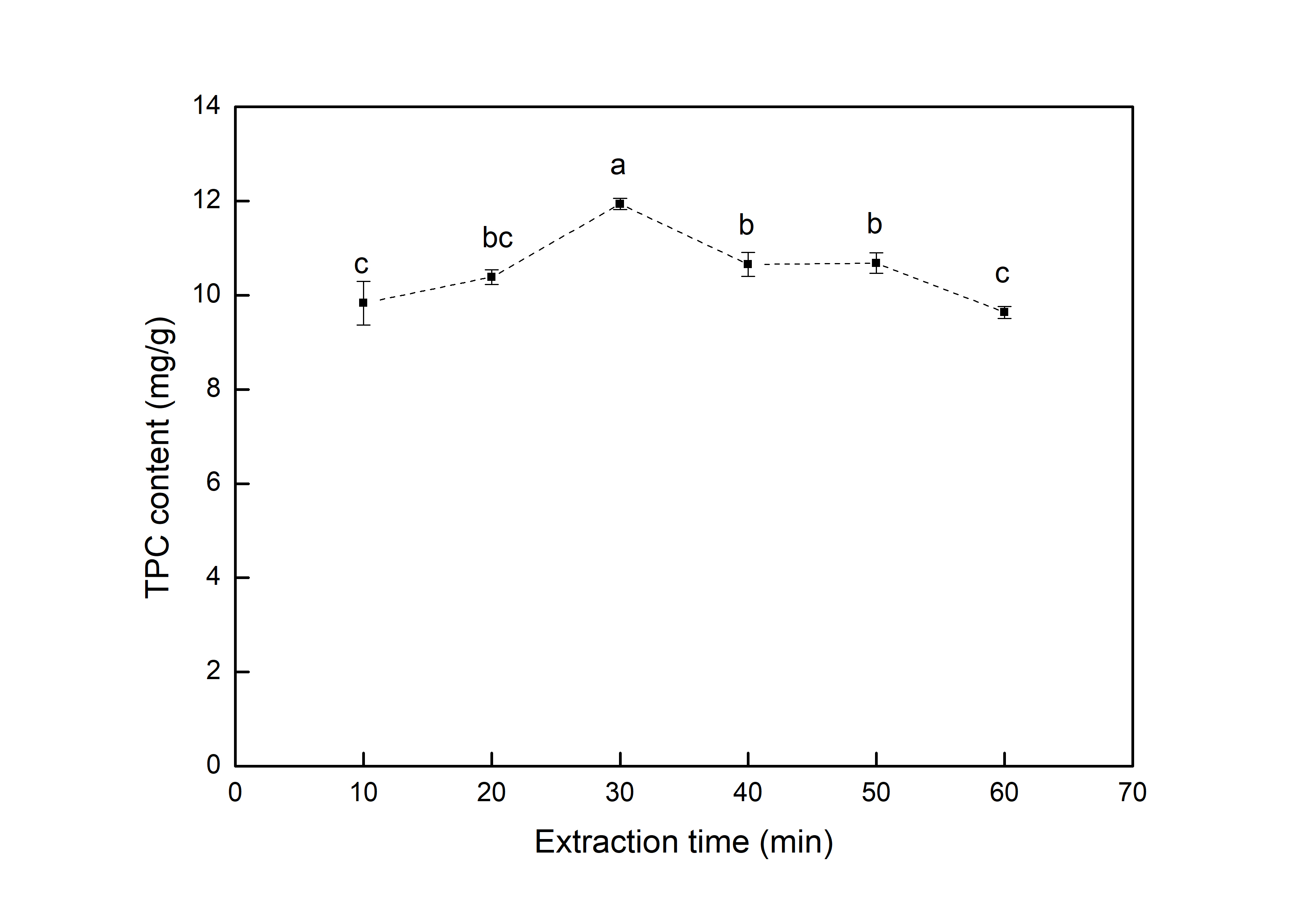 Note: Values followed by different letters are significantly different by the Tukey test (p ≤ 0.05).
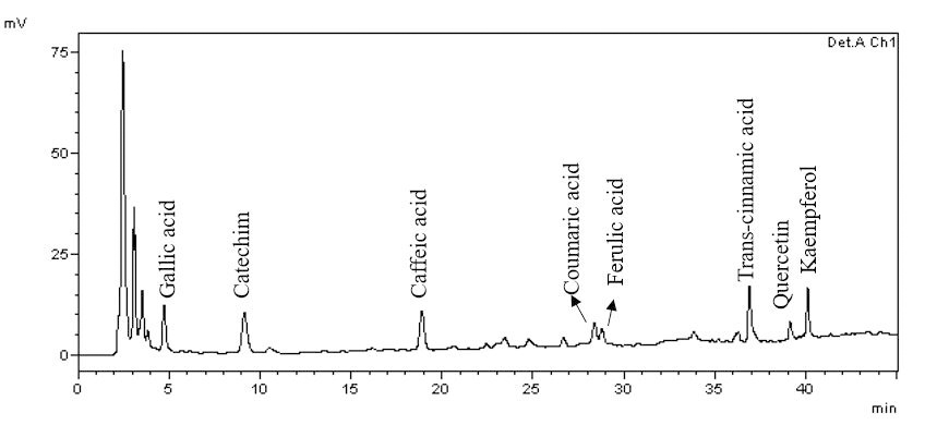 Note: Extraction conditions: 125 rpm, 60 °C, 1:30 g mL-1. Det A: UV detector at 280 nm.