In vitro evaluation of the antioxidant, antibacterial and cytotoxic activities of Osteospermum moniliferum L. leaf and stem extracts
Abstract
Osteospermum moniliferum is an unexplored shrub with regard to its pharmacological properties. In this study, the antioxidant potential of O. moniliferum was evaluated using the 2,2 diphenyl-1-picrylhydrazyl (DPPH) and ferric-reducing antioxidant power (FRAP) assays. The DPPH assay indicated that the stem methanolic extract demonstrated the most pronounced scavenging capability (83.16%) at the minimal concentration of 15 mg/mL, with IC50 values recorded at 5.08 mg/mL. The IC50 values obtained from the FRAP assay were suboptimal for the leaf extracts (>1000 µg/mL), whereas the stem extracts exhibited remarkable activity, with values of 2.75 mg/mL for methanol, 3.52 mg/mL for chloroform, and 4.49 mg/mL for hexane extracts. The cytotoxicity was evaluated against three human cell lines, viz. breast adenocarcinoma (MCF-7), embryonic kidney (HEK293) and lung carcinoma (A549), using the (MTT) assay. Moderate activity was observed for the leaf (44.66±0.09 µg/µL) and stem (47.37±0.08 µg/µL) hexane extracts for the HEK293 cell line. In contrast, stem methanol, leaf methanol, leaf chloroform and stem chloroform extracts had low cytotoxic activity against the MCF-7 cell line. All extracts exhibited promising antibacterial activity against Staphylococcus aureus, Escherichia coli, and Pseudomonas aeruginosa. These findings could potentially contribute to the advancement of naturally derived compounds used in healthcare.
Key words
Antioxidant; cytotoxicity; antibacterial; Osteospemum moniliferum; Asteraceae
INTRODUCTION
The rate at which infectious diseases spread worldwide is very worrying (Soyemi, 2021). The emergence of antibiotic-resistant bacterial strains presents a significant global health challenge, highlighting the urgent need to identify alternative antimicrobial agents (Sharaf et al. 2021). Antimicrobial compounds are utilized as antibiotics to treat infections in the human body, but they can have various adverse effects, including an increase in reactive oxygen species (ROS) (Raut et al. 2019, Shaikh et al. 2019). ROS are highly detrimental to human health and well-being, playing a role in the development of cancer (Raut et al. 2019, Kim et al. 2016). They may also worsen potential medical issues (Parham et al. 2019, Panieri & Santoro, 2016). Plant extracts with antibacterial properties offer a promising solution to combatting resistant bacteria and reducing the reliance on conventional antibiotics.
Cancer is a fatal disease spreading rapidly and is a growing global issue due to the lack of widespread and comprehensive early detection tools and appropriate therapy (Khalil et al. 2018). Cancer remains one of the leading causes of death globally, responsible for approximately ten million deaths in 2020 (Ferlay et al. 2020). This disease is characterized by uncontrollable or irreversible cell proliferation, resulting in the development of cancerous tumours with the potential to spread through metastatic lesions (Greenwell & Rahman, 2015). By 2030, it is projected that there will be 26 million new cancer cases and 17 million cancer-related deaths annually (Thun et al. 2009). According to Charles-Okhe et al. (2022), women are more prone to developing breast, colorectal, lung, cervical, and thyroid cancers, while men are more likely to develop lung, prostate, colorectal, stomach, and liver cancers. Lung cancer is reported to be responsible for 18.4% of all cancer-related deaths worldwide, followed by breast (11.6%), stomach (8.2%), liver (8.2%), prostate (7.1%) and colorectal cancers (6.1%) (de Martel et al. 2020). Various combinations of surgery, chemotherapy, and radiation therapy are commonly employed in cancer treatment (Mathan et al. 2022). Conventional therapies and cytotoxic medications with synthetic bases are often expensive for the underprivileged, and chemotherapies have major side effects (Senapati et al. 2018). Therefore, medicinal plants and plant-based therapies play an essential role in primary healthcare, particularly for rural populations, in preventing and treating a broad spectrum of diseases, including cancer (Koche et al. 2016).
Herbal plants have been found to contain a variety of phytochemicals with antioxidant properties, and their application in cancer treatment has demonstrated a reduction in toxicities and adverse side effects (Bhuyan et al. 2017, Dutt et al. 2019). More than 60% of anticancer medications, including vinblastine (Catharanthus roseus), vincristine (Catharanthus roseus), camptothecin (Camptotheca acuminita), taxol (Taxus brevifolia), podophyllotoxin, and combretastatin (Combretum caffrum), are said to be derived from natural sources (Cragg & Newman 2005, Ediriweera et al. 2019). These compounds combat reactive oxygen species (ROS), resulting in less oxidative stress, improved immune function and increased longevity (Tan et al. 2018). Moreover, medicinal plants contain a broad array of naturally occurring antioxidants that may help alleviate oxidative stress and its harmful effects on human health by scavenging or stabilizing free radicals (Matteo & Esposito, 2003, Soltanian et al. 2020). Therefore, medicinal plants are a rich source of herbal medicines for preventing and treating various ailments.
Asteraceae members are widely distributed worldwide and have been utilized in traditional medicine since ancient times (Achika et al. 2014, Rolnik & Olas 2021). Plants from this family are known to contain various metabolites used in cancer treatment (Rolnik & Olas 2021). The species within Asteraceae contains multiple phytochemical compounds, including polyphenols, phenolic acids, flavonoids, sesquiterpene lactones, saponins and lignans. In our previous study, we analyzed the leaves and stems of O. moniliferum, and identified the presence of various phytochemical compounds with potential biological activities (Mhlomi et al. 2025). While sufficient scientific reports confirm the ethnopharmacological importance of plants from this family, the antioxidant, antibacterial and anticancer properties of O. moniliferum remain vague, and its pharmacological effectiveness needs to be further investigated. Therefore, the current study used established protocols to determine the antioxidant, cytotoxic and antibacterial activities of leaf and stem extracts of O. moniliferum.
MATERIALS AND METHODS
Plant collection
Osteospermum moniliferum was collected within the University of KwaZulu-Natal (UKZN), Westville campus (29°49’7.21” S; 30°56’50.45” E). The plant was authenticated by Dr Syd Ramdhani (Curator of the Ward Herbarium), and the voucher specimen (NU0094285) was deposited in the Bews Herbarium, Pietermaritzburg campus of the UKZN. The leaves and stem bark materials were washed, dried, pulverised and stored in an airtight container for further use.
Preparation and extraction of plant material
The fresh plant materials (leaves and stems) were collected and then air-dried at room temperature for four weeks. A mill was used to grind the dried plant materials into powder. Approximately 10 g of each powdered material was subjected to a sequential extraction using a reflux apparatus. One hundred millilitre solvent was used for each three-hour sample extraction and the process was repeated thrice. The solvents utilised were hexane, chloroform, and methanol solvent, in order of increasing polarity. Extracts were filtered using Whatman No. 1 (Whatman Limited, Maidstone, UK) filter paper. Extracts were allowed to dry in the dark at room temperature (23±2°C). Dried extracts were subsequently preserved in airtight, appropriately labelled containers at a temperature of 4°C until required for subsequent applications. The percentage yield of the extract was calculated utilizing the formula presented below:
In vitro antioxidant activity
2,2 diphenyl-2-picrylhydrazyl (DPPH) radical scavenging activity
The free radical scavenging activity was performed using 2,2 diphenyl-1-picrylhydrazyl (DPPH) using a modified method by Braca et al. (2003). Five different concentrations of the investigated plant extracts (15, 30, 60, 120 and 240 µg/mL) were prepared in methanol. DPPH (0.3 mM) was prepared using 99% methanol. Briefly, 50 µL of 0.3 mM prepared DPPH solution was added to 100 µL of each extract. The mixtures were thoroughly mixed before incubating in the dark for 30 min at 23°C. The colourimetric observation from purple to yellow suggested that the extracts displayed scavenging activity. The absorbance was quantified at a wavelength of 517 nm employing a Synergy HTX Multi-mode reader (BioTek Instruments Inc., Winooski, VT, USA). Ascorbic acid served as the calibration standard. The efficacy of the extracts in scavenging DPPH radicals was ascertained through the application of the following equation:
Where Abs control is the absorbance of DPPH and methanol, and Abs sample is the absorbance of the DPPH radical + sample or standard.
The experiments were performed in triplicates. The half maximum inhibitory (IC50) of the extracts was calculated using a plot of the percentage of DPPH free radical inhibition against the extract concentration.
Ferric-reducing antioxidant power (FRAP) assay
The antioxidant capacity of the extracts was assessed in accordance with the methodology established by Akwu et al. (2019). In summary, 50 µL of each extract was combined with 50 µL of 0.2 M sodium phosphate buffer (pH 6.6) at standard concentrations (15, 30, 60, 120, and 240 µg/mL) and 100 µL of 1% potassium ferricyanide. The resultant mixture was incubated for 30 minutes at a temperature of 50°C. The reaction was terminated by the addition of 50 µL of 10% trichloroacetic acid, 50 µL of distilled water, and 10 µL of 0.1% iron (III) chloride (FeCl3). The mixture was permitted to stand for ten minutes, after which the absorbance was quantified at 700 nm utilizing a Synergy HTX Multi-mode reader (BioTek Instruments Inc., Winooski, VT, USA). Gallic acid served as the positive control in this experiment. The results were expressed as a percentage of the absorbance of the extracts relative to that of gallic acid using the formula provided below:
Anticancer activity
Cell culture
The cytotoxic properties of the extracts were evaluated against three distinct human cell lines, specifically breast adenocarcinoma (MCF-7), human embryonic kidney (HEK293), and human lung carcinoma (A549), which were generously provided by the Department of Biotechnology at Durban University of Technology. These cell lines were cultured to confluency in a 25 cm² tissue culture flask and maintained in Dulbecco’s Eagle’s minimum essential medium (DMEM) at a temperature of 37 °C with a CO2 concentration of 5% within a HEPA Class 100 steri-Cult CO2 incubator (Thermo-Electron Corporation, Waltham, Massachusetts, USA). The DMEM was further supplemented with 10% (v/v) gamma-irradiated Fetal Bovine Serum (FBS) and 1% antibiotics (10,000 units/mL penicillin, 10,000 U/mL streptomycin). Subsequently, the cells underwent trypsinization, were seeded into a 96-well plate, and incubated for a duration of 24 hours at 37 °C to facilitate cellular adhesion. The growth medium was subsequently refreshed with entirely new medium (DMEM + 10% FBS + 1% antibiotics) for cell preparation.
The potential cytotoxic effects of the extracts derived from the leaves and stems of O. moniliferum were investigated utilizing the 3-(4.5 dimethylthiazol-2yl)-2,5-diphenyltetrazolium bromide (MTT) assay across three cell lines. The MTT assay was executed in accordance with the methodology delineated by Dwarka et al. (2017). The cells were administered 50 µL of the six extracts at varying concentrations ranging from 7.5 to 1000 µg/mL and subsequently cultured at 37 °C for 24 hours. Camptothecin was employed as a positive control in this experimental setup. Post-incubation, MTT reagent (20 µL, 5 mg/mL) was introduced to each well and incubated at 37 °C for a period of 4 hours. Following this incubation, the MTT medium was aspirated and replaced with 100 µL of dimethyl-sulphoxide (DMSO). The absorbance of the resulting solution was measured at 570 nm utilizing a Multiscan Go microplate reader (Thermo Scientific, Waltham, MA, USA). The percentage viability of the cells was subsequently computed using the following formula:
In-vitro antibacterial activity
Preparation of bacterial strains
Various American Type Culture Collection (ATCC) reference bacterial strains, specifically Staphylococcus aureus (ATCC 43300), Escherichia coli (ATCC 25922), and Pseudomonas aeruginosa (ATCC 27853), were generously supplied by Professor Johnson Lin from the Department of Microbiology at the University of KwaZulu-Natal. These bacterial strains were preserved in a 75% glycerol solution at a temperature of -80°C. A loopful of each bacterial strain was cultivated overnight in a test tube shaker for a duration of 24 hours at a temperature of 36°C. Subsequently, each bacterial culture (inoculum) was further diluted with sterile nutrient broth to achieve an optical density (OD) ranging from 0.08 to 0.1 at 625 nm, as measured by a UV-visible spectrophotometer, resulting in a final concentration of approximately 1 × 10⁸ to 1 × 10⁹ bacterial cells per milliliter.
Antibacterial bioassay
The antibacterial activity of extracts (hexane, chloroform, and methanol) derived from the leaves and stem bark of O. moniliferum was assessed utilizing the agar well diffusion methodology delineated by Akwu et al. (2019). Different concentrations of the extracts (0.625, 1.25, 2.5, 5 and 10 mg/mL) were prepared briefly in dimethyl sulfoxide (DMSO). Mueller-Hinton Agar (MHA) was used to make agar plates. Agar was poured into Petri plates at room temperature and was allowed to solidify. The bacterial cultures were swabbed onto the plates using sterile cotton swabs. The wells were punched using a sterile cork borer with a 6-mm diameter. After that, 100 µL of each prepared extract concentration was pipetted into the wells. The remaining cups on each plate were filled with a standardised dose (10 μg/mL) of streptomycin and gentamycin as controls for strains of Gram-positive and Gram-negative bacteria, respectively. The plates were incubated at 36°C to promote the growth of bacterial colonies. The antibacterial activity of the plates was determined by measuring the diameter of the bacterial zone of inhibition (mm) after 18 to 24 h of incubation. The activity of each extract was tested in triplicates.
Statistical Analysis
Minitab statistical software version 17.0 was used for the analysis. Data were analyzed in triplicate (n=3) and were presented as means ± standard deviation. A One-Way Analysis of Variance (ANOVA) was used to see if the means of the different groups differed significantly. Tukey’s test for pairwise comparison and mean separation was additionally conducted. Values were considered significant when p< 0.05.
RESULTS AND DISCUSSION
In vitro antioxidant activity
Presently, tracking the interest in the antioxidant activity of plant or food extracts is a popular way to investigate the potential therapeutic properties (Ghani et al. 2019). Antioxidant activity protects tissues and organs from the harmful effects of free radicals, which may lead to disease development (e.g., cancer and cardiovascular diseases) (Pfingstgraf et al. 2021, Aljaja et al. 2021). The ability of several types of samples to scavenge free radicals has been evaluated using the free radical compound DPPH (Kusuma et al. 2014). The DPPH radical scavenging assay constitutes a highly sensitive evaluation of antioxidant capacity, which is predicated on hydrogen donation, substrate polarity, and/or radical scavenging mechanisms. This methodology is extensively recognized as a parameter for assessing a plant extract’s proficiency in neutralizing free radicals and pertains to the capacity for hydrogen atom or electron donation that is independent of any enzymatic processes (Mileva et al. 2014). The antioxidant capacity of hexane, chloroform and methanol extracts from leaves and stem of O. moniliferum were evaluated using DPPH radical scavenging activity and FRAP (Figures 1 and 2). The extracts of O. moniliferum demonstrated a remarkable in vitro DPPH radical scavenging activity in a dose dependent manner (Figure 1). The methanol stem extract showed higher scavenging activity (%) than other extracts. At the lowest concentration (15 µg/mL), the stem methanol extract exhibited the highest scavenging activity (83.16%), followed by the leaf hexane (72.07%), leaf chloroform (72.02%) and leaf methanol (53.14%) extracts. Whereas at the highest concentration (240 µg/mL), stem methanol (91.15 %) had the highest scavenging activity, followed by leaf methanol (89.97%) and stem hexane (83.97%). Overall, the methanolic stem extract showed a greater percentage of scavenging activities. The percentage scavenging activities of the extract were significantly lower than that of the standard (ascorbic acid). Additionally, the IC50 values of the extracts were higher than the IC50 value of the ascorbic acid (Table I). These results are in accordance with the result from the study by Muthoni Guchu et al. (2020) who demonstrated the antioxidant capacity of three plant species. The stem extract had better IC50 values compared to the leaf extracts, with stem hexane (4.84 µg/mL), methanol (5.08 µg/mL) and chloroform (6.08 µg/mL) extracts displaying better activity than the leaves. The leaf methanolic extract showed a higher antioxidant activity with an IC50 value of 16.21 µg/mL than other leaf extracts. This function may also be linked to the presence of alkaloids, flavonoids and phenolic compounds that are present in plants. Osteospermum monilifera stem and leaves are relevant as a potential organic source of antioxidants and can be used to treat diseases caused by free radicals due to their lower reported IC50 values. The results from the current study are comparable to those reported by Kommidi et al. (2014). The antioxidant activity of leaves, stems, and root fractions were investigated using DPPH. The results were concentration-dependent, with the methanol stem fraction having a higher scavenging activity (94 µg/mL) at the highest concentration of 250% compared to other extracts. The ability of an extract to effectively reduce a substance is known to reflect any antioxidant properties a plant may have (Do et al. 2014). Due to the complexity of the compounds contained in extracts, Mwihia (2017) reaffirmed that it is necessary to assess the antioxidant potential of the plant using at least two techniques.
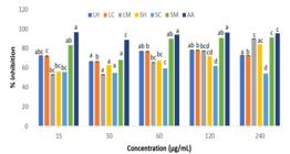
In vitro antioxidant activity (% inhibition DPPH) using leaf hexane (LH), leaf chloroform (LC), leaf methanol (LM), stem hexane (SH), stem chloroform (SC), stem methanol (SM) extracts of O.moniliferum. AA: Ascorbic acid. Superscripts indicate a significant difference (P<0.05) between the means. Sets of bars with different superscripts are significantly different.
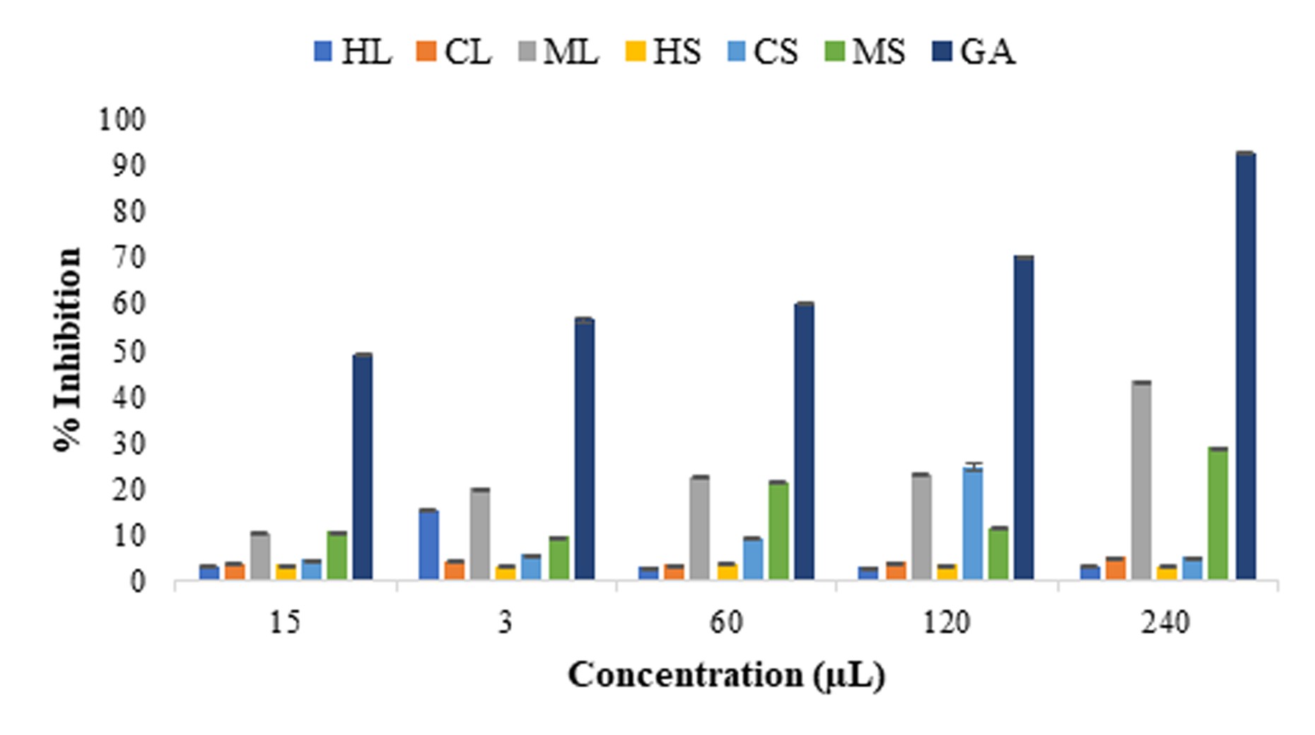
The FRAP assay was another technique for measuring antioxidant activity. This approach was based on the ability of the analyte to reduce the ferric ion (Fe3+) to ferrous ion (Fe2+) (Adesanoye & Farombi 2014). Hence, the formation of Fe2+ can be examined at 700 nm absorbance capacity. Absorbance increases at this wavelength indicate an increase in reducing power (Muthoni Guchu et al. 2020). The FRAP assay revealed low percentage inhibition across extracts and doses ranging from 2.98% to 43.27% compared to the control, which displayed a percentage inhibition of 49.02% at 15 µg/mL and 90.52% at 240 µg/mL (Figure 2). Overall, the methanolic extracts showed a better percentage of inhibition than other extracts and increased with increased concentration. The IC50 values of the FRAP assay were weak for the leaf extracts (> 1000 µg/mL), while the stem extracts displayed excellent activity (2.75 µg/mL for methanol, 3.52 µg/mL for chloroform and 4.49 µg/mL for hexane) (Table I). The low IC50 observed in stem extracts suggests the extracts have high antioxidant activity. Similar results from the DPPH and FRAP experiments indicated that the stem extracts had a greater antioxidant potential than the leaf extracts. The variation in the antioxidant capacity between the previous research and the current investigation is most likely due to differences in the geographic location and environmental conditions (soil and climate) of the plant sample or the type of plant organ used (Sari et al. 2020, Raju & Rao 2021).
IC50 values of antioxidant activities of different extracts from leaves and stem of Osteospermum moniliferum. NA = not applicable.
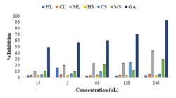
In vitro, antioxidant activity (% inhibition FRAP) of crude extracts from Osteospemum moniliferum leaves and stem. HL – hexane leaves, CL – chloroform leaves, ML – methanol leaves, HS – hexane stem, CS –chloroform stem, MS – methanol stem, GA – gallic acid.
Cytotoxicity activity
There exists a pressing necessity to formulate anticancer therapeutics that specifically target malignant cells while preserving the integrity of normal cells (Sidambaram et al. 2011). Various plant species and their bioactive components play a crucial role in impeding cancer progression and in the innovation of novel, clinically effective anticancer pharmaceuticals (Kaur et al. 2011, Iqbal et al. 2017). For the first time, the cytotoxic activity was evaluated utilizing hexane, chloroform, and methanol extracts derived from the leaves and stems of O. moniliferum against the MCF-7, HEK293, and A549 cell lines. The impact of the extracts at varying concentrations (7.81–1000 µg/mL) was ascertained through the MTT assay. This assay is founded on the capacity of the mitochondrial dehydrogenase enzyme from viable cells to cleave the tetrazolium rings of the pale yellow MTT, thereby generating dark blue formazan crystals that exhibit limited permeability to cellular membranes, culminating in their accumulation within healthy cells (Maposa et al. 2022). MTT reduction correlates with cellular protein and viable cell number (Braissant et al. 2020). To establish the anticancer capabilities of the extract, it must be toxic to the MCF-7 or A549 cancer cells and exhibit a minor reactivity to HEK293, with additional IC50 data. Cytotoxic action in malignant cells increases as the corresponding IC50 values decrease. The results of the MTT assay are graphically illustrated in Figure 3. The percentage viability of the cancer cells (MCF-7 and A594) exhibited a marked decline at elevated concentrations. The results showed that at the lowest concentration (7.8125 µg/µL), MCF-7 cells treated with the leaf extracted in chloroform were the most sensitive (95.23%), while at the highest concentration (1000 µg/µL), stems extracted in hexane displayed substantial cell sensitivity (23.21%). Our results suggested that the extracts had more cytotoxic potential against the A549 cancer cell line than against MCF-7.

In vitro cytotoxicity (% cell survival) of leaf hexane (LH), leaf chloroform (LC), leaf methanol (LM), stem hexane (SH); stem chloroform (SC); stem methanol (SM); control (Camptothecin). (A) MCF-7, (B) HEK293, and (C) A549 cell lines. Superscripts indicate a significant difference (P<0.05) between the means. Bars with different superscripts are significantly different.
The results showed that tested extracts exhibited different potencies of cytotoxicity activity against two cancer cell lines at eight different concentrations (Figure 3). Substantial cytotoxic effects were displayed by stem methanol extracts (0.49 ± 0.45 µg/µL) for the HEK293 cell line and leaf hexane (9.30 ± 0.55 µg/µL), stem hexane (8.78 ± 0.05 µg/µL), leaf methanol (28.37 ± 0.17 µg/µL) and stem methanol extracts (33.60 ± 0.16 µg/µL) for the A549 cell line (Table II). Moderate activity was observed for leaf (44.66 ± 0.09 µg/µL) and stem (47.37 ± 0.08 µg/µL) extracted in hexane for the HEK293 cell line, whereas the stem methanol, leaf methanol, leaf chloroform and stem chloroform extracts exhibited weak cytotoxic activity against the MCF-7 cell line with IC50 values of 634.4 ± 0.22 µg/µL, 412.6 ± 0.24 µg/µL, 219.4 ± 0.13 µg/µL and 106.2 ± 0.10 µg/µL, respectively.
Leaves and flower aqueous extracts of Chrysanthemum suppressed the proliferation of MCF-7 cell lines in a dose-dependent manner at concentrations greater than 25 µg/mL (Murayama et al. 2013). Lee et al. (2014) on the anticancer potential of some Korean Chrysanthemum species (C. boreale, C. indicum and C. morifolium) found that the methanolic extracts inhibited MCF-7 cell lines quite well, with cell viability ranging from 47–63% doses as 200 mg/mL. It was shown that treatment with 200 mg/mL of C. morifolium reduced cell viability by 49%. A study by Luo et al. (2010) revealed that Vernonia amygdalina extracts inhibited the proliferation of MCF-7 and BT549. Furthermore, Kouamé et al. (2013) investigated the cytotoxicity of Chromolaena odorata extracts in MCF-7, MDBAMB-468, CAL51 Lewis lung carcinoma (LLC) and HL-60 cancer cell lines using different solvents. The investigation indicated that the n-hexane leaf extract demonstrated inhibitory effects on MCF-7, MDAMB-468, and CAL51 breast cancer cell lines, whereas the ethanolic leaf extract exhibited suppression of LLC and HL-60 (Kouamé et al. 2013). Plants from the Asteraceae are known to contain various bioactive compounds such as alkaloids, tannins, flavonoids, glycosides, phenolic, saponins and quinines that contribute to their biological activities (Bessada et al. 2015, Koc et al. 2015). For instance, it has been documented that a prominent compound present in members of the Asteraceae family is a sesquiterpene lactone. Numerous pharmacological attributes, including antimicrobial, anticancer, and anti-inflammatory effects, have been recognized for sesquiterpene lactones. (Ravi & Bedi 2004, Guzman et al. 2005, Dhyani et al. 2022). Notably, while the methanolic stem extract exhibited higher antioxidant capacities than the other tested extracts (of the present study), the extract exhibited a lower cytotoxic effect against MCF-7 (Figure 3a). This anomaly may have been due to variations in the extracts’ phytoconstituents and the cell’s sensitivity towards the extract.
Zone of inhibition (mm) of hexane extracts of Osteospermum moniliferum at different concentrations.
Antibacterial activity
According to Maddila & Hamalathe (2017), bacterial resistance to most current antibiotics has been observed, increasing antibacterial resistance globally. To combat bacterial resistance, the pharmaceutical industry and new biotechnology business are expanding their search for a novel antibacterial agent (de Souza et al. 2023). Osteospermum moniliferum leaf and stem extracts were tested for antibacterial activity against three pathogenic bacterial strains at five concentrations (0.625, 1.25, 2.5, 5, and 10 mg/mL). The bacteria included Gram-positive bacteria (Staphylococcus aureus) and Gram-negative bacteria (Escherichia coli, Pseudomonas aeruginosa). From the results, it is evident that the zone of inhibition increased with increasing concentration. All extracts for both leaves and stem inhibited the growth of the bacterial strains (E. coli, S. aureus and P. aeruginosa) but had minor antibacterial activity when compared to the controls (antibiotics). The results revealed that the maximum zone of inhibition ranged from 6.2– 9.33 mm for both Gram-negative and Gram-positive bacterial strains in hexane extracts (Table III). The zone of inhibition of the leaf and stem extracts at 10 mg/mL against E. coli was significantly higher when compared to the control (p<0.05) (Table IV, V). The zone of inhibition at other doses was lower in relation to the antibiotic controls. It was noted that the zone of inhibition was dose-dependent in all extracts.
Zone of inhibition (mm) of chloroform extracts of Osteospermum moniliferum at different concentrations.
Zone of inhibition (mm) of methanol extracts of Osteospermum moniliferum at different concentrations.
Secondary metabolites found in plant cells allow the leaf and stem extracts to inhibit bacterial growth (Nurtjahja et al. 2013). While comparing the efficacy extracts against Gram-positive and Gram-negative bacterial strains, it was observed that the extracts were more effective on the Gram-positive bacterial strain. These results are in accordance with those of Bibiso et al. (2021). Staphylococcus aureus showed higher susceptibility to the extracts than the other tested strains. Gram-negative bacteria were expected to be more resistant as they have been reported to be more resistant than Gram-positive to antibiotics (Palombo & Semple 2001). Gram-negative bacteria present significant medical challenges due to their robust outer membrane, which is constituted of an asymmetric bilayer formed by phospholipids and lipopolysaccharides, thereby functioning as a barrier against a plethora of environmental substances, as well as detergents and antibiotics (such as penicillin) that would typically compromise the integrity of the peptidoglycans within their (inner) cell membrane. In comparison to hexane and chloroform, methanol extracts of O. moniliferum displayed superior inhibition against the chosen bacterial strains. It can be inferred that methanol serves as an efficacious extracting solvent, proficiently solubilizing phytocompounds endowed with antibacterial properties (Sanwaral & Sushil 2013). Additional studies yielding analogous results further corroborate this conclusion (Seleshe & Kang, 2019, Verma & Verma 2019, Cudjoe et al. 2020). The ability of plant extracts to inhibit distinct bacterial strains supports their use as anti-infection agents and points to their antimicrobial action. Furthermore, the formation of inhibitory zones against both Gram-negative and Gram-positive bacteria demonstrates their applicability for a wide range of applications.
According to Saglam & Arar (2003), pure compounds and medicinal plant extracts have demonstrated antibacterial activity in both in vitro and in vivo studies. Therefore, phytochemicals such as phenols, tannins, saponins, and flavonoids are known to be biologically active and thus may have played a role in the antibacterial effects of the plant (Dhatwalia et al. 2021). Plant extracts with well-known antibacterial properties might be used for therapeutic purposes, which could increase the use of herbal treatments for microbial illness (Unuofin et al. 2018). The observed moderate antibacterial efficacy implies that these plant species may represent a viable source of natural compounds and antibiotics exhibiting varying degrees of antiseptic properties. Strains of Staphylococcus aureus have been associated with a range of medical conditions, including superficial skin and soft tissue infections, infective endocarditis, ditis, osteomyelitis, bacteremia, lethal pneumonia, as well as instances of foodborne illness (Tong et al. 2015, Guo et al. 2020). The results derived from this investigation indicate that extracts from the leaves and stems of O. moniliferum have the potential to be utilized in the treatment of infections attributable to this particular bacterial strain.
CONCLUSIONS
This study evaluated the antioxidant, cytotoxic, and antibacterial properties of O. moniliferum to determine its potential therapeutic value. Each extract exhibited substantial antioxidant properties to varying degrees, suggesting that they may protect against free radicals and oxidative damage associated with various diseases. The correlation between radical-scavenging agents in the extracts and their cytotoxicity in cancer cells may facilitate data screening for natural compounds with cytotoxic potential. Indeed, the extracts demonstrated a cytotoxic effect against cancerous cells in vitro, which may be attributed to the antioxidant activity of the species’ various components. Furthermore, the extracts were effective against both Gram-negative and Gram-positive bacteria. To the best of our knowledge, this is the first study on the cytotoxicity and antioxidant potential of this plant. Considering that Osteospermum is an underutilized genus of Asteraceae, further scientific investigation, such as antidiabetic assays, is recommended.
Acknowledgements
Support by the National Research Foundation (NRF), South Africa, is greatly appreciated. The authors declare no conflicts of interest.
References
- ACHIKA JI, ARTHUR DE, GERALD I & ADEDAYO A. 2014. A review on the phytoconstituents and related medicinal properties of plants in the Asteraceae family. IOSR J Appl Chem 7: 1-8.
- AKWU N, NAIDOO Y, SINGH M, NUNDKUMAR N & LIN J. 2019. Phytochemical screening, in vitro evaluation of the antimicrobial, antioxidant and cytotoxicity potentials of Grewia lasiocarpa E. Mey. ex Harv. South Afr J Botany 123: 180-192.
- ALJAJA MK, LAHDO R & KITAZ A. 2021. phytochemical study and protective effect investigation against oxidative damage in human erythrocytes of Syrian Asteraceae plants. Bulletin of Pharmaceutical Sciences. Assiut 44: 323-338.
- BESSADA SM, BARREIRA JC & OLIVEIRA MBP. 2015. Asteraceae species with most prominent bioactivity and their potential applications: A review. Industrial Crops and Products 76: 604-615.
- BHUYAN DJ, VUONG QV, BOND DR, CHALMERS AC, VAN ALTENA IA, BOWYER MC & SCARLETT CJ. 2017. Exploring the least studied Australian eucalypt genera: Corymbia and Angophora for phytochemicals with anticancer activity against pancreatic malignancies. Chem Biodivers 14: e1600291.
- BIBISO M, ANZA M & ALEMAYEHU B. 2021. Antibacterial and antioxidant activity of Cirsium Englerianum (Asteraceae), an endemic plant to Ethiopia. Res J Pharmacogn 8: 5-12.
- BRACA A, FICO G, MORELLI I, DE SIMONE F, TOMÈ F & DE TOMMASI N. 2003. Antioxidant and free radical scavenging activity of flavonol glycosides from different Aconitum species. J Ethnopharmacol 86: 63-67.
- BRAISSANT O, ASTASOV-FRAUENHOFFER M, WALTIMO T & BONKAT G. 2020. A review of methods to determine viability, vitality, and metabolic rates in microbiology. Front Microbiol 11: 547458.
- CHARLES-OKHE O, ODENIYI MA, FAKEYE TO, OGBOLE OO & AKINLEYE TE & ADENIJI AJ. 2022. Cytotoxic activity of crude extracts and fractions of African peach nauclea latifolia (smith) stem bark on two cancer cell lines. Phytomed Plus 2: 100212.
- CRAGG GM & NEWMAN DJ. 2005. Plants as a source of anti-cancer agents. J Ethnopharmacolog 100: 72-79.
-
CUDJOE E, DONU D, OKONU RE, AMPONSAH JA & AMOAH LE. 2020. The in vitro antiplasmodial activities of aqueous extracts of selected Ghanaian herbal plants. J Parasitol Res 2020: doi: 10.1155/2020/5041919.
» https://doi.org/10.1155/2020/5041919 - DE MARTEL C, GEORGES D, BRAY F, FERLAY J & CLIFFORD GM. 2020. Global burden of cancer attributable to infections in 2018: a worldwide incidence analysis. The Lancet Global Health 8: e180e190.
- DE SOUZA GHDA, ROSSATO L, DE OLIVEIRA AR & SIMIONATTO S. 2023. Antimicrobial peptides against polymyxin-resistant Klebsiella pneumoniae: a patent review. World J Microbiol Biotechnol 39: 86.
- DHATWALIA J, KUMARI A, VERMA R, UPADHYAY N, GULERIA I, LAL S, THAKUR S, GUDETA K, KUMAR V & CHAO JC-J. 2021. Phytochemistry, pharmacology, and nutraceutical profile of Carissa species: An updated review. Molecules 26: 7010.
- DHYANI P, SATI P, SHARMA E, ATTRI DC, BAHUKHANDI A, TYNYBEKOV B, SZOPA A, SHARIFI-RAD J, CALINA D & SULERIA HA. 2022. Sesquiterpenoid lactones as potential anti-cancer agents: an update on molecular mechanisms and recent studies. Cancer Cell Int 22: 1-18.
- DO QD, ANGKAWIJAYA AE, TRAN-NGUYEN PL, HUYNH LH, SOETAREDJO FE, ISMADJI S & JU YH., 2014. Effect of extraction solvent on total phenol content, total flavonoid content, and antioxidant activity of Limnophila aromatica J Food Drug Analysis 22: 296-302.
- DUTT R GARG V, KHATRI N & MADAN AK. 2019. Phytochemicals in anticancer drug development. Anticanc Agents Med Chemi 19: 172-183.
- DWARKA D, THAVER V, NAIDU M, KOORBANALLY NA, & BAIJNATH H. 2017. In vitro chemo-preventative activity of Strelitzia nicolai aril extract containing bilirubin. African Journal of Traditional, Complementary and Alternative Medicines 14: 147-156.
- EDIRIWEERA MK, TENNEKOON KH & SAMARAKOON SR. 2019. In vitro assays and techniques utilized in anticancer drug discovery. J Appl Toxicol 39: 38-71.
- FERLAY J, ERVIK M, LAM F, COLOMBET M, MERY L, PIÑEROS M, ZNAOR A, SOERJOMATARAM I & BRAY F. 2020. Observatory: “Cancer Today”. Lyon International Agency Research on Cancer.
- GHANI MA, BARRIL C, BEDGOOD JR DR & PRENZLER PD. 2019. Development of a method suitable for high-throughput screening to measure antioxidant activity in a linoleic acid emulsion. Antioxidants 8: 366.
- GUO L, GONG S, WANG Y, SUN Q, DUO K & FEI P. 2020. Antibacterial activity of olive oil polyphenol extract against Salmonella Typhimurium and Staphylococcus aureus: Possible Mechanisms. Foodborne Pathog Dis 17: 396-403.
- GUZMAN ML, ROSSI RM, KARNISCHKY L, LI X, PETERSON DR, HOWARD DS & JORDAN CT. 2005. The sesquiterpene lactone parthenolide induces apoptosis of human acute myelogenous leukemia stem and progenitor cells. Blood 105: 4163-4169.
- GREENWELL M & RAHMAN PKSM. 2015. Medicinal plants: their use in anticancer treatment. Int J Pharm Sci Res 6: 4103.
- IQBAL J, ABBASI BA, MAHMOOD T, KANWAL S, ALI B, SHAH SA & KHALIL AT. 2017. Plant-derived anticancer agents: A green anticancer approach. Asian Pacific J Trop Biomed 7:1129-1150.
- KAUR R, SINGH J, SINGH G & KAUR H. 2011. Anticancer plants: A review. J Natural Prod Plant Res 1: 131-136.
- KHALIL N, ASHOUR M, SINGAB AN & SALAMA O. 2018. Bioassay guided fractionation and cytotoxic activity of Daucus carota var. boissieri. Future J Pharm Sci 4: 4-17.
- KIM S-H, KIM K-Y, YU S-N, PARK S-G, YU H-S, SEO Y-K & AHN S-C. 2016. Monensin induces PC-3 prostate cancer cell apoptosis via ROS production and Ca2+ homeostasis disruption. Anticancer Research 36: 5835-5843.
- KOC S, ISGOR BS, ISGOR YG, SHOMALI MOGHADDAM N & YILDIRIM O. 2015. The potential medicinal value of plants from Asteraceae family with antioxidant defense enzymes as biological targets. Pharmaceut Biolo 53: 746-751.
- KOCHE D, SHIRSAT R & KAWALE M. 2016. An overview of major classes of phytochemicals: their types and role in disease prevention. Hislopia J 9: 0976-2124.
- KOMMIDI DR, KANDAPPA HR, MOODLEY B, KOORBANALLY NA & GOVENDER P. 2014. Bio evaluation of different crude extracts of Chrysanthimoides monilifera subsp. rotundata. Turk J Biochem 39.
- KOUAMÉ PBK, JACQUES C, BEDI G, SILVESTRE V, LOQUET D, BARILLÉ-NION S, ROBINS RJ & TEA I. 2013. Phytochemicals isolated from leaves of Chromolaena odorata: Impact on viability and clonogenicity of cancer cell lines. Phytother Res 27: 835-840.
- KUSUMA IW, ARUNG ET & KIM YU. 2014. Antimicrobial and antioxidant properties of medicinal plants used by the Bentian tribe from Indonesia. Food Sci Human Welln 3: 191-196.
- LEE JH, CHOI EJ, PARK HS & KIM GH. 2014. Evaluation of Compositae sp. plants for antioxidant activity, anti-inflammatory, anticancer and antiadipogenic activity in vitro Food Agric Immunol 25: 104-118.
- LUO X, OYUGI DA, LIN C, IZEVBIGIE EB & LEE KS. 2010. Isolation and characterization of the antibreast carcinoma cell growth components of Vernonia amygdalina extracts. Exper Biol Med 235: 1472-1478.
- MADDILA S & HEMALATHA KPJ. 2017. Phytochemical screening and in vitro antimicrobial properties of crude leaf extracts of Wrightia tinctoria R. Br. Int J Curr Microbiol Appl Sci 6: 707-720.
- MAPOSA S, OTUNOLA GA & AFOLAYAN AJ. 2022. Cytotoxicity evaluation of Moringa oleifera (M. oleifera) Lam. flowers on human liver cancer cell line and A. salina (Brine shrimp) models. Int J Pharmaceut Sci Res 13: 4909-4917.
- MATHAN SV, RAJPUT M & SINGH RP. 2022. Chemotherapy and radiation therapy for cancer. Understanding Cancer: 217-236.
- MATTEO V & ESPOSITO E. 2003. Biochemical and therapeutic effects of antioxidants in the treatment of Alzheimer’s disease, Parkinson’s disease, and amyotrophic lateral sclerosis. Current Drug Targets CNS & Neurological Disorders 2: 95-107.
- MHLOMI Y, NAIDOO Y, NAIDOO KK, DEWIR YH, EL-HENDAWY S, ALSHAHRANI TS & PATIL R. 2025. Phytochemical constituents of tickberry (Osteospermum moniliferum L.) Leaf and stem extracts. Appl Ecol Environ Res 23: 3839-3860.
- MILEVA M, KUSOVSKI VK, KRASTEV DS, DOBREVA AM & GALABOV AS. 2014. Chemical composition, in vitro antiradical and antimicrobial activities of Bulgarian Rosa alba L. essential oil against some oral pathogens. Int J Curr Microbiol Appl Sci 3:11-20.
- MURAYAMA K, SAWA T, KOGA H, YAMAGUCHI S, KUMAZOE M, OTSUKA T, MIYAZAKI Y, TACHIBANA H & YAMADA K. 2013. Anticancer effect of water extracts prepared from Chrysanthemum flowers and leaves Shiranui Himekiku. Nippon Shokuhin Kagaku Kogaku Kaishi 60: 80-86.
- MUTHONI GUCHU B, MACHOCHO AKO, MWIHIA SK & NGUGI MP. 2020. In vitro antioxidant activities of methanolic extracts of Caesalpinia volkensii Harms., Vernonia lasiopus O. Hoffm., and Acacia hockii De Wild. Evidence-Based Complementary and Alternative Medicine 1: 3586268.
- MWIHIA SK. 2017. In vitro antibacterial and antioxidant activities of methanolic and dichloromethanolic seed extracts of Kenyan Annona squamosa Linn. Kenyatta University, Nairobi, Kenya.
- NURTJAHJA K, KELANA TB, SURYANTO D, PRIYANI N, RIO G, PUTRA DP & ARBAIN D. 2013. Antimicrobial activity of endemic herbs from Tangkahan Conservation Forest north Sumatera to bacteria and yeast. HAYATI J Biosci 20:177-181.
- PALOMBO EA & SEMPLE SJ. 2001. Antibacterial activity of traditional Australian medicinal plants. J Ethnopharmacol 77:151-157.
- PANIERI E & SANTORO M. 2016. ROS homeostasis and metabolism: a dangerous liason in cancer cells. Cell Death and Disease 7: e2253-e2253.
- PARHAM S, WICAKSONO DH & NUR H. 2019. A proposed mechanism of action of textile/Al2O3–TiO2 bimetal oxide nanocomposite as an antimicrobial agent. J Textile Inst 110: 791-798.
- PFINGSTGRAF IO, TAULESCU M, POP RM, ORĂSAN R, VLASE L, UIFALEAN A, TODEA D, ALEXESCU T, TOMA C & PÂRVU AE. 2021. Protective effects of taraxacum officinale L.(dandelion) root extract in experimental acute on chronic liver failure. Antioxidants 10: 504.
- RAJU M & RAO YV. 2021. Study of Catalase, Protease, Antioxidant and Antimicrobial Activities of Tabernaemontana divaricata Latex. J Med Plants By-prod 10: 61-68.
- RAUT PK, KIM S-H, CHOI DY, JEONG G-S & PARK P-H. 2019. Growth of breast cancer cells by leptin is mediated via activation of the inflammasome: Critical roles of estrogen receptor signaling and reactive oxygen species production. Biochem Pharmacol 161: 73-88.
- RAVI R & BEDI A. 2004. NF-κB in cancer - A friend turned foe. Drug resistance updates 7: 53-67.
- ROLNIK A & OLAS B. 2021. The plants of the Asteraceae family are agents in the protection of human health. Int J Mol Sci 22: 3009.
- SAGLAM H & ARAR G. 2003. Cytotoxic activity and quality control determinations on Chelidonium majus Fitoterapia 74: 127-129.
- SANWARAL A & SUSHIL K. 2013. Antibacterial activity of Mangifera indica leaves against drugresistant bacterial strain. Int J Adv Res 1: 82-86.
- SARI R, CONTERNO P, DA SILVA LD, DE LIMA VA, OLDONI TLC, THOMÉ GR & CARPES ST. 2020. Extraction of phenolic compounds from Tabernaemontana catharinensis leaves and their effect on oxidative stress markers in diabetic rats. Molecules 25: 2391.
- SELESHE S & KANG SN. 2019. In vitro, antimicrobial activity of different solvent extracts from Moringa stenopetala leaves. Prevent Nutr Food Sci 24: 70.
- SENAPATI S, MAHANTA AK, KUMAR S & MAITI P. 2018. Controlled drug delivery vehicles for cancer treatment and their performance. Signal Transduction and Targeted Therapy 3: 7.
- SHAIKH S, NAZAM N, RIZVI SMD, AHMAD K, BAIG MH, LEE EJ & CHOI I. 2019. Mechanistic insights into the antimicrobial actions of metallic nanoparticles and their implications for multidrug resistance. Int J Mol Sci 20: 2468.
- SHARAF MH, EL-SHERBINY GM, MOGHANNEM SA, ABDELMONEM M, ELSEHEMY IA, METWALY AM & KALABA MH. 2021. New combination approaches to combat methicillin-resistant Staphylococcus aureus (MRSA). Sci Rep 11: 4240.
- SIDAMBARAM RR, DINESH MG & JAYALAKSHMI ET. 2011. An in vitro study of cytotoxic activity of Euphorbia hirta on Hep2 cells of human epithelioma of larynx. Int J Pharm Pharmaceut Sci 3: 3.
- SOLTANIAN S, SHEIKHBAHAEI M, MIRTADZADINI M & KHANDANI BK. 2020. Evaluation of anticancer, antioxidant and antibacterial properties of methanol extract of three Acantholimon Boiss. Species. Avicenna J Phytomed 10: 641.
- SOYEMI T. 2021. Neglect of common infectious disease outbreaks during the COVID-19 pandemic: an impending crisis in Nigeria? African J Clinic Exper Microbiol 22: 113-116.
- TAN BL, NORHAIZAN ME, LIEW WPP & SULAIMAN RAHMAN H. 2018. Antioxidant and oxidative stress: a mutual interplay in age-related diseases. Front Pharmacol 9: 1162.
- THUN MJ, DELANCEY JO, CENTRE MM, JEMAL A & WARD EM. 2009. The global burden of cancer: priorities for prevention. Carcinogenesis 31: 100-110.
- TONG SY, DAVIS JS, EICHENBERGER E, HOLLAND TL, & FOWLER JR VG. 2015. Staphylococcus aureus infections: epidemiology, pathophysiology, clinical manifestations, and management. Clinic Microbiol Rev 28: 603-661.
- UNUOFIN JO, OTUNOLA GA & AFOLAYAN AJ. 2018. Polyphenolic content, antioxidant and antimicrobial activities of Vernonia mespilifolia Less. used in folk medicine in the Eastern Cape Province, South Africa. J Evidence-Based Integr Med 23: 2515690X18773990.
- VERMA P & VERMA RK. 2019. Succession of culturable microbes on rhizosphere soil of iron ore mined overburden dump in Dalli Rajhara, Durg, Chhattisgarh, India. Plant Archives 19: 1179-1189.
Edited by
-
Handling editor
Vasco Azevedo
Data and materials supporting the results or analyses presented in the article are available upon reasonable request.