Analysis of the antimicrobial activity of propolis: A narrative review of in-vitro studies
Abstract
Propolis, harvested by bees from local and seasonal flora, exhibits a diverse chemical composition renowned for its antimicrobial, antioxidant, and anti-inflammatory attributes. This positions it as a promising ally in combatting antimicrobial resistance. This narrative review aims to provide a comprehensive investigation of in vitro studies investigating propolis’ antimicrobial efficacy. Employing the Preferred Reporting Items for Systematic Reviews and Meta-Analyses (PRISMA) methodology, we conducted searches in PubMed, SciELO, and Lilacs databases. Articles published between 2011 and 2023, in English, Spanish, or Portuguese, and focusing on propolis’ in vitro antimicrobial activity against human pathogens were included using descriptors. Out of 291 initially identified articles, 34 were meticulously chosen post-application of exclusion criteria. Despite originating from diverse regions, propolis consistently demonstrates a rich profile abundant in fundamental phenolic compounds and flavonoids. The synergy of its constituent elements’ leads to significant antibacterial significant antibacterial and antifungal effects via distinct mechanisms. Appreciating the varied chemical compositions of propolis is imperative for optimizing its therapeutic potential. Its remarkable antibacterial and antifungal attributes, alongside standardized testing methodologies and exploration of synergistic combinations with conventional antibiotics or essential oils, present compelling strategies for effectively addressing antimicrobial resistance.
Key words
antimicrobial activity; bees; natural products; propolis
INTRODUCTION
The indiscriminate use of antibiotics has led to an increase in resistance mechanisms among various microorganisms, creating a need for new alternatives to combat infections, especially those of bacterial and fungal origin (Brinkac et al. 2017). This looming crisis is projected to reach catastrophic proportions by 2050, with antimicrobial resistance causing an alarming increase in morbidity and accounting for an estimated 10 million deaths annually (Bassetti et al. 2017). Beyond its devastating human toll, antimicrobial resistance also exacts a heavy toll on the global economy. In response to this pressing issue, natural products have emerged as a promising tool for combating antimicrobial resistance. These natural products offer a diverse array of chemical compounds and functional substances that have demonstrated their effectiveness against resistant microorganisms. In essence, they represent a vital alternative in the ongoing battle against this global health threat (Ye et al. 2020).
Considering the urgency of the situation, the World Health Organization (WHO) has recognized the significance of Traditional, Complementary, and Integrative Medicine (TCIM) practices. These approaches, rooted in ancestral experiences, offer valuable insights into disease prevention and recovery. Consequently, an increasing number of countries are adopting TCIM as part of their healthcare strategies. By 2023 it was reported that 170 countries have implemented public policies that regulate and promote the use of TCIM, aligning with WHO’s recommendations (WHO 2023). These approaches often incorporate natural substances, harnessing the therapeutic potential of botanicals, minerals, and other naturally occurring compounds. The use of herbal medicine, dietary supplements, and traditional practices, alongside conventional medical interventions, aims to address the physical, mental, and emotional aspects of health.
Propolis is a complex mixture of resinous and balsamic substances, of varied consistency, texture, and coloration, collected by Apis mellifera bees or stingless bee species, from various parts of plants, such as floral buds, shoots, and resinous exudates, in the vicinity of the apiary. The bees also add salivary secretions, wax, and pollen, which accounts for the variation in its coloration, texture, and consistency (Park et al. 2002, Funari et al. 2016). They use this substance to protect themselves against microorganisms and insects, applying it in thin layers on the internal walls of the hive to seal cracks, repair and strengthen honeycombs, protect the entrance of the hive, and in the preparation of aseptic places for the laying of eggs by the queen bee and in the mummification of invading insects (Bankova et al. 2000). In Brazil, products derived from beekeeping, like propolis, have been officially incorporated into the national policy of integrative and complementary practices (Política Nacional de Práticas Integrativas e Complementares [PNPIC]). Moreover, such policy is specifically tailored for users of the Brazilian Unified Health System (Sistema Único de Saúde [SUS]) throughout the entire national territory (Brasil 2018).
The chemical composition of propolis reflects the flora used by bees (Elnakady et al. 2017), and can vary according to regional seasonality, which can influence its potential for action and its physical, chemical, and biological properties (Sforcin et al. 2000, Castro et al. 2007). However, it is usually composed of 50% plant resin and balsam, 30% wax, 10% essential and aromatic oils, 5% pollen, and 5% other various substances, including organic residues (Alaerjani et al. 2022). Considered one of the most heterogeneous natural mixtures, it is believed that more than 300 different substances have been identified and/or characterized from different propolis samples (Alanazi et al. 2021). In general, propolis can contain flavonoids, modified carboxylic acids, higher hydrocarbons, alcohols, aromatic acids, higher fatty acids typical of waxes and their esters, ketones, flavones and flavanols, flavanones, chalcones and dihydrochalcones, terpenoids, steroids, amino acids, sugars, lignans, vitamins (A, B1, B2, B6, C, E, and PP) and minerals (sodium, potassium, magnesium, barium, strontium, cadmium, lead, copper, manganese, iron, calcium, vanadium, silicon, aluminum, nickel, zinc, chromium, titanium, silver, molybdenum, and cobalt) (Bankova et al. 2000, dos Santos Pereira et al. 2002, Kalogeropoulos et al. 2009, Wozniak et al. 2023).
Brazilian propolis has been classified into twelve classes according to Park et al. (2002), with a diverse range of colors. In the South region alone, five types are identified, displaying hues from yellow to dark brown and greenish-brown. The Northeast region contributes six distinct types, including reddish-brown and various shades of yellow and green. The Southeast is known primarily for its brown, greenish-brown, and green varieties. Additionally, a thirteenth type originating from mangroves in the states of Sergipe, Alagoas, Paraíba, Pernambuco, and Bahia was introduced in 2007. Among these varieties, the green, red, and brown types stand out as the most researched and economically significant, particularly for their notable biological activities and export value, especially to Asian countries (Figueiredo-Rinhel et al. 2013).
Some components are present in all propolis samples, while others are specific to propolis derived from plant species (Vargas et al. 2004, Bobis 2022). Notably, propolis exhibits a diverse array of beneficial properties, including anti-inflammatory, antibacterial, immunomodulatory, antioxidant, antidiabetic, antiprotozoal, antihypertensive, antihepatotoxic, and antiviral effects (Przybyłek & Karpiński 2019, Salamanca-Grosso et al. 2022, Bobis 2022).
Notably, propolis’ antibacterial properties have garnered significant attention in research, showing effectiveness against diverse bacteria (Salamanca-Grosso et al. 2022) and fungi, including yeast (Bezerra et al. 2020) and filamentous fungi (Wolska & Antosik 2023). Given the substantial number of research highlighting the advantages of propolis, it is crucial to conduct a comprehensive assessment of its potential as an antimicrobial agent. Therefore, the primary aim of this systematic review is to offer a comprehensive overview of studies assessing the in vitro antimicrobial activity of propolis.
MATERIALS AND METHODS
Search strategy
This research adhered to the Preferred Reporting Items for Systematic Reviews and Meta-Analyses (PRISMA) methodology, as outlined by Page et al. (2022). The identification of relevant studies was conducted through comprehensive searches on databases such as National Library of Medicine (PubMed), Online Scientific Electronic Library (SciELO), and Latin American and Caribbean Literature on Health Sciences (Lilacs). Utilizing the Patient, Intervention, Comparison and Outcome (PICO) strategy, the central research question was formulated as follows: Among studies assessing the in vitro antimicrobial activity of propolis, what consensus exists regarding its efficacy against both bacteria and fungi? These searches employed search terms in alignment with the Descritores em Ciências da Saúde/Medical Subject Headings (DeCS/MeSH) terminology (BIREME 2022). The search was conducted by associating the following descriptors using the “all fields” search option: (própolis e antimicrobianos) or (própolis e antibacterianos) or (própolis e antifúngicos) in Portuguese, (propolis and anti-infective agents) or (propolis and anti-bacterial agents) or (propolis and antifungal agents) in English, and (Própole y antimicrobianos) or (Própole y antibacterianos) or (Própole y Antifúngicos) in Spanish.
Eligibility criteria
It was included articles in English, Spanish, and Portuguese, with the primary objective of examining the in vitro antimicrobial activity of propolis against human pathogens. The selected articles encompassed publications from the years 2011 to 2023.
Study selection process and data extraction
Data extraction from each database was conducted by GLL, DCL, and PP. Subsequently, initial selection of studies was performed by S.R.A. through reading the titles and abstracts. Review of study integrity and accuracy concerning the proposed objectives was undertaken by R.C.S.V. The final selection of studies was made by J.K.B and R.H.P. An Excel™ spreadsheet was employed to systematically collect the following information: author’s name, year of publication, country of origin, research objectives, microorganism strain type, inoculum concentration, extract type utilized, susceptibility testing method, results for the treated group, and concluding remarks.
RESULTS
During the initial phase of study searching, descriptors and their combinations were utilized as detailed in Figure 1. In total, it was identified 291 studies, with 269 sourced from the three databases and an additional 22 studies obtained from other resources. As depicted in Figure 1, during the identification phase, duplicate studies were excluded, resulting in a total of 139 articles. Subsequent evaluation of titles and abstracts led to the removal of 62 studies that did not examine the antimicrobial activity of propolis. We proceeded to a full reading of 77 articles. Among these, 37 articles were excluded for not meeting the criteria as in vitro investigations, and an additional 5 articles were excluded as they assessed the antimicrobial activity of propolis on non-infectious agents for humans. After the eligibility assessment, 34 relevant studies were selected for the narrative review.

Table I presents a succinct overview of the characteristics of the selected studies. The systematic review encloses studies primarily differing in terms of the microorganisms under investigation, with 15 articles examining various bacterial genera, 6 articles focusing on different fungal species, and 7 studies addressing both bacteria and fungi.
Characteristics of the selected studies: study location, microorganism, type of propolis extract used, method employed, and principal results.
DISCUSSION
Propolis composition: regional variability, extraction methods, and implications for biological activity
The biological properties of propolis tend to vary according to the specific region from which propolis samples are obtained in each zone and continent worldwide. Despite this variability, propolis generally exhibits some common characteristics, which have been utilized to classify it into distinct classes. Propolis can typically be categorized into two primary types: the Baccharis-type (Brazilian-type) and the Poplar-type (European-type). The Brazilian-type propolis is abundant in derivatives of p-coumaric acid, such as artepillin C, (E)-3-prenyl 4-(dihydrocinnamoyl-oxy)-cinnamic acid, dihydrokaempferol, 4-hydroxy-3-prenylbenzoic acid, and plicatin B. Conversely, the European type propolis, collected not only in Europe but also in China and other countries, is characterized by its richness in flavonoids and phenolic acid esters, notably pinocembrin, pinobanksin, galangin, chrysin, and caffeic acid phenethyl ester, as it primarily originates from bud exudates of the populus species (Benhanifia et al. 2014, Ristivojević et al. 2016).
The chemical composition of propolis is influenced not only by the geographic region of collection but also by various other factors, such as its botanical origin (Bucio-Villalobos & Martínez-Jaime 2017, Salamanca-Grosso et al. 2022, Sa-Eed et al. 2023), the time of collection, the species of bee, and the solvent used for extraction (Rufatto et al. 2018). Consequently, it is important to interpret findings from specific studies with caution, as they may not fully represent the propolis composition across entire countries or regions.
For instance, South American propolis shows considerable diversity. Mexican propolis from Cuautitlán Izcalli and Michoacán was found to have a higher percentage of flavonoids, particularly pinocembrin, while propolis from Sonora was rich in pinocembrin, xanthomicrol, chrysin, and galangin (Rodríguez Pérez et al. 2020). Chilean propolis from La Araucanía exhibited notable levels of apigenin, pinocembrin, quercetin, and caffeic acid phenethyl ester (CAPE) (Veloz et al. 2019). Argentinean propolis from Bahia Blanca in Buenos Aires province contained abundant phenolic compounds (Cibanal et al. 2019). Brazilian propolis from Mato Grosso do Sul had high concentrations of kaurenoic acid, benzoic acid, cinnamic acid, and 3-phenyl-p-coumaric acid (Campos et al. 2015), whereas propolis from Paraná was particularly rich in flavonoids like quercetin (Barbosa et al. 2014).
Similarly, findings from other regions emphasize the variability of propolis composition. Algerian propolis from Tiaret and Tlemcen contains various phenolic compounds, including caffeic acid, ferulic acid, apigenin, pinobanksin, CAPE, chrysin, pinocembrin, and galangin (Benhanifia et al. 2014). Serbian propolis is often categorized into two types, orange and blue, with the orange variety predominating and containing phenolics such as caffeic acid, galangin, quercetin, and chrysin (Ristivojević et al. 2016). German propolis contains several acids, including benzoic acid, cinnamic acid, and salicylic acid, while Irish propolis is rich in flavonoids (chrysin, galangin, and pinocembrin) as well as nonacosane, pentacosane, and alpha-bisabolol. Czech propolis is dominated by phenyl carboxylic acids (caffeic acid, cinnamic acid, and benzoic acid) along with flavonoids (Al-Ani et al. 2018).
Distinctive compositions are observed in Spanish and Brazilian propolis, influenced by their specific botanical sources. Spanish propolis, likely derived from olive trees, contains compounds like vanillic acid, 1-acetoxypinoresinol, elenolic acid mono-aldehyde, and oleuropein-aglycone di-aldehyde (Navarro-Pérez et al. 2021, Fernández-Calderón et al. 2021). Brazilian red propolis, primarily from Dalbergia ecastophyllum plants, has a unique profile including (3S)-vestitol, medicarpin, benzoic acid, and liquiritigenin (Rufatto et al. 2018). Additionally, Silva et al. (2019) identified red flavonones, xanthones, pentacyclic triterpenoids, and leucoanthocyanidins in red propolis. Figure 2 shows the main components of propolis with antimicrobial activity and the number of regions where each was identified, and Figure 3 illustrates the molecular structures of these compounds.

Main antimicrobial components of propolis and the frequency of their identification across different regions. The bar chart highlights the diversity of propolis compounds with antibacterial and antifungal properties, reflecting regional variation in chemical composition.

Molecular structures of various compounds found in propolis with known antibacterial activity.
Considering these findings, we acknowledge the limitations inherent in characterizing propolis composition from limited studies within each region. Propolis composition can vary even within the same country due to local botanical sources and environmental factors. We suggest that further studies covering multiple localities within each country would be beneficial to establish a more representative understanding of regional propolis compositions.
Interestingly, despite the common association of Apis mellifera with propolis production, Barreiras et al. (2020) and Campos et al. (2023) conducted studies on propolis produced by the stingless bee Tetragonisca fiebrigi collected from the state of São Paulo and Mato Grosso do Sul, Brazil, respectively. According to Campos et al. (2023), the propolis from these bees exhibited phenolic compounds, alcohol, and terpenes as its major class compounds. Later, the same group of authors (Campos et al. 2023) investigated propolis produced by Melipona quadrifasciata anthidioides and Scaptotrigona depilis bees in Brazil. These propolis samples, designated as EEP-M and EEP-S, respectively, displayed a composition rich in phenolic compounds, flavonoids, and triterpenes. This highlights the diversity of propolis compositions influenced by different bee species and their foraging habits and underscores the potential of exploring propolis from various bee sources as an option in the search for new therapeutic alternatives.
Furthermore, mineral ion content varies across different geographic regions, as highlighted by Moghim et al. (2021) in Iranian propolis, which contains silver, mercury, copper, manganese, iron, and vanadium, and by Salamanca-Grosso et al. (2022) in Colombian propolis, which contains calcium, iron, potassium, magnesium, and manganese, reflecting the influence of environmental factors on propolis composition.
The yield of phenolic compounds in propolis is closely linked to the extraction method used, as shown in the Figure 4. Hydroalcoholic extraction (Silva et al. 2012, Moncayo Luján et al. 2018, Cibanal et al. 2019, Petruzzi et al. 2020, Barreiras et al. 2020), and ultrasound-assisted extraction (Moncayo Luján et al. 2018, Sokolonski et al. 2021) methods are particularly effective, resulting in the highest relative yields of phenolic compounds, with ultrasound pre-treatment enhancing specific compounds such as isoflavones and kaempferol (Sokolonski et al. 2021). In contrast, aqueous extraction produces significantly lower yields of active phenolic components (Bucio-Villalobos & Martínez-Jaime 2017). Ethanolic extraction remains widely utilized (Bastos et al. 2011, Alves Ferreira Bastos et al. 2011, Probst et al. 2011, Dantas de Almeida et al. 2012, Barbosa et al. 2014, Benhanifia et al. 2014, Campos et al. 2015, Joya et al. 2017, Maureira et al. 2017, Al-Ani et al. 2018, Veloz et al. 2019, Silva et al. 2019, Correa et al. 2020, Rodríguez-Perez et al. 2020, Mateo Aldama et al. 2020, Navarro-Pérez et al. 2021, Fernández-Calderón et al. 2021, Papp et al. 2021, Moghim et al. 2021, Sokolonski et al. 2021, Salamanca-Grosso et al. 2022, Campos et al. 2023), offering a moderate yield, while methanolic extraction is efficient for certain polar compounds, such as flavonoid aglycones (Rivera-Yañez et al. 2022). Additionally, chloroform and ethyl acetate were noted to effectively fractionate antimicrobial components in specific studies (Sa-Eed et al. 2023).
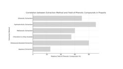
Correlation between various extraction methods and the relative yield of phenolic compounds in propolis. Hydroalcoholic and ultrasound-assisted extractions demonstrate superior effectiveness, yielding the highest concentrations of phenolic compounds and highlighting their potential for optimizing bioactive compound recovery. In contrast, aqueous extraction results in significantly lower yields, underscoring the importance of selecting an appropriate extraction method to maximize the therapeutic potential of propolis.
Nevertheless, the quality of propolis can be compromised by impurities, including beeswax residue, water, ash, and mechanical contaminants such as remnants of vegetation or bees, dyes, and vegetable powder (Checalla-Collatupa & Sánchez-Tito 2021, Rivera-Yañez et al. 2022). The beeswax content is subject to various factors such as collection time, beekeeper handling, propolis-producing bee species, and extraction method. Higher humidity levels may encourage mold and yeast growth, while ash content is linked to metallic contaminants like lead, iron, and copper (Rodríguez-Perez et al. 2020). In Brazil, the “Identity Regulation and Quality of Propolis Extract” outlined in Instruction n° 03, dated January 19, 2001, seeks to define the identity of Brazilian propolis extract and set minimum quality standards for both domestic and international trade (Rufatto et al. 2018). Among these standards is the requirement for a minimum phenolic compound content, set at 0.50% (w/w).
Antibacterial activity of propolis
The antibacterial impact of propolis varies between Gram-negative and Gram-positive bacteria, primarily owing to distinctions in the structure and arrangement of the cell wall (Bastos et al. 2011, Silva et al. 2012, Barbosa et al. 2014, Campos et al. 2015, Ristivojević et al. 2016, Barreiras et al. 2020, Sa-Eed et al. 2023) although the action is more pronounced against Gram positive bacteria (Alves Ferreira Bastos et al. 2011, Silva et al. 2012, 2019, Benhanifia et al. 2014, Rufatto et al. 2018, Bucio-Villalobos & Martínez-Jaime 2017, Rodríguez-Perez et al. 2020, Barreiras et al. 2020, Salamanca-Grosso et al. 2022, Sa-Eed et al. 2023). The antibacterial activity may be attributed to multiple targets, with various constituents (phenolic compounds, diterpenes, and flavonoids) acting synergistically, as shown in Figure 5.

Antibacterial action of propolis based on the findings of Silva et al. (2012), Barbosa et al. (2014), Campos et al. (2015), Ristivojević et al. (2016), Moncayo Luján et al. (2018), Veloz et al. (2019), Petruzzi et al. (2020), Rodríguez-Pérez et al. (2020), Navarro-Pérez et al. (2021), Sa-Eed et al. (2023), and Campos et al. (2023). Propolis components, particularly phenolic compounds, interact with bacterial cell structures through multiple mechanisms. Key actions include damage to the plasma membrane, DNA gyrase inhibition, disruption of ATP synthesis, and interference with protein synthesis. These combined actions hinder bacterial growth and demonstrate the potential of propolis as an antibacterial agent.
In the context of biofilms, which shield microorganisms within a matrix of polymeric substances, including those formed by Streptococcus mutans, apigenin and pinocembrin found in propolis are suggested to disrupt the activity of glucosyltransferase (GTF) enzymes (Veloz et al. 2019, Checalla-Collatupa & Sánchez-Tito 2021). The GTF C enzyme is responsible for producing both water-insoluble and soluble glucans (dextran), while GTF D exclusively synthesizes water-soluble glucans. These glucans, particularly the water-insoluble mutan, are believed to play a crucial role in promoting adhesion and colonization of the organism. Moreover, they are thought to mediate protection against antimicrobial agents and confer resistance to toxic compounds (Thurnheer et al. 2006). Furthermore, a study by Navarro-Pérez et al. (2021) demonstrated that the Spanish propolis extract, characterized by high polyphenol content, especially in the flavonoid class, and containing unique compounds as vanillic acid, 1-acetoxypinoresinol, p-HPEA-EA, and 3,4-DHPEA-EDA, inhibited the formation of mono and double species biofilms of S. mutans and Streptococcus sanguinis. Additionally, scanning electron microscopy (SEM) images revealed significant clustering among S. sanguinis cells, while S. mutans produced relatively homogeneous biofilms (Navarro-Pérez et al. 2021).
Antifungal activity of propolis
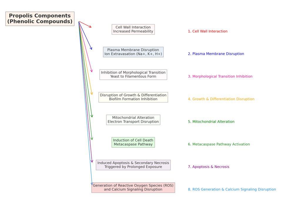
The antifungal mechanisms of propolis phenolic compounds are detailed in Figure 6. These compounds interfere with fungal cell structures—targeting the cell wall, plasma membrane, and mitochondria—leading to increased permeability, ion leakage, apoptosis, and secondary necrosis, ultimately inhibiting fungal growth (Campos et al. 2015, Rufatto et al. 2018, Correa et al. 2020, Petruzzi et al. 2020, Rodríguez-Perez et al. 2020, Sokolonski et al. 2021, Fernández-Calderón et al. 2021, Mateo Aldama et al. 2020, Campos et al. 2023, Rivera-Yañez et al. 2022).

Antifungal action of propolis based on the studies of Campos et al. (2015), Rufatto et al. (2018), Correa et al. (2020), Petruzzi et al. (2020), Rodríguez-Pérez et al. (2020), Sokolonski et al. (2021), Fernández-Calderón et al. (2021), Mateo Aldama et al. (2020), Campos et al. (2023), and Rivera-Yañez et al. (2022). Propolis components, particularly phenolic compounds, exert antifungal effects by interacting with the cell wall, increasing its permeability; disrupting the plasma membrane, causing ion leakage (Na+, K+, H+); and altering mitochondrial function, which interferes with the electron transport chain and energy production. Prolonged exposure can also induce apoptosis, followed by secondary necrosis. These combined actions underscore propolis’s potential as a powerful antifungal agent.
The antifungal efficacy of propolis primarily arises from its phenolic compounds (Correa et al. 2020, Sokolonski et al. 2021, Campos et al. 2023). These compounds thwart fungal growth by interacting with the cell wall (Rufatto et al. 2018, Correa et al. 2020, Rodríguez-Perez et al. 2020, Rivera-Yañez et al. 2022), and plasma membrane (Fernández-Calderón et al. 2021), leading to heightened permeability and produces extravasation of sodium, potassium and hydrogen ions, causing the fungus to die (Campos et al. 2015, Correa et al. 2020, Rodríguez-Perez et al. 2020, Mateo Aldama et al. 2020, Campos et al. 2023). At the mitochondrial level, they induce alterations in the electron transport chain, ultimately triggering apoptosis (Campos et al. 2015, Petruzzi et al. 2020, Correa et al. 2020); prolonged exposure exacerbates this effect, resulting in secondary necrosis (Petruzzi et al. 2020). It also prevents the morphological transition from yeast to filamentous forms (Fernández-Calderón et al. 2021, Rivera-Yañez et al. 2022), impedes cell growth and biofilm formation (Correa et al. 2020, Rodríguez-Perez et al. 2020, Sokolonski et al. 2021), and activates the metacaspase pathway for cell death. Furthermore, propolis stimulates reactive oxygen species (ROS) generation (Correa et al. 2020, Fernández-Calderón et al. 2021, Rivera-Yañez et al. 2022), and interferes with calcium signaling pathways (Campos et al. 2015, Rivera-Yañez et al. 2022), enhancing its antifungal efficacy.
When Cryptococcus neoformans was exposed to Mexican propolis, containing 22.7% ± 0.007 of phenolic compounds, notable changes in morphology were observed, including a decrease in size (average of 2.186 µm), the formation of pores (average of 0.651 µm), and invaginations on the surface of the yeast (Mateo Aldama et al. 2020).
Alterations in the cell surface of Candida albicans were observed, attributed to the interaction between sulfhydryl compounds of yeast with propolis, mimicking imidazoles (Mateo Aldama et al. 2020). Furthermore, compounds such as caffeic acid, flavonoids and phenolic esters from Mexican propolis (specifically from the Cuautitlán region) showed inhibition of the germ tube and changes in the cell wall of C. albicans (Mateo Aldama et al. 2020). Rivera-Yañez et al. (2022) reported that Mexican propolis exhibited inhibition of germ tube formation within a range of 19 µg/mL to 1290 µg/mL. The ability of propolis samples to inhibit germ tube growth suggests promising potential in the search for novel antifungal agents. This process marks the initial stage for C. albicans to initiate pathogenic behavior, limiting their full range of pathogenic mechanisms.
Rivera-Yañez et al. (2022) also highlighted that pinocembrin demonstrates antifungal activity, which is associated with a decrease in the cell surface hydrophobicity of C. albicans as well as decreased ALS3 (Agglutinin-Like Sequence, gene encoding cell-wall proteins), and ACT1 (ACTin, gene that encoding actin) messenger Ribonucleic Acid (mRNA) levels. Indeed, the chemical configuration of pinocembrin, particularly the hydroxy group at the five position, the ketone group at the four position, and the six-member condensed with benzene, plays a crucial role in its inhibitory effect on biofilm formation. Furthermore, Andrade-Pavón et al. (2022) have reported that naringenin and naringin exhibit an anti-Candida effect and can recognize and binding to enzymes such as topoisomerase II in Candida sp. It is worth noting that topoisomerase II plays a crucial role in the dynamics of gene expression, as this enzyme is necessary for the correct unwinding and compaction of DNA, and therefore, for its replication (Andrade-Pavón et al. 2022). Kaempferol has also been reported as an inhibitor of fluconazole-resistant C. albicans strains, with Minimal Inhibitory Concentration (MIC) ranging from 128–256 μg/mL. The mechanism underlying the inhibitory effect of this flavonoid is associated with a reduction in the expression of the Candida Drug Resistance genes (CDR1/CDR2, genes encoding membrane transport proteins of the ABC transporter) and Multidrug Resistance (MDR1) genes, which are involved in fluconazole resistance in C. albicans by controlling the positive regulation of the multidrug efflux pump (Shao et al. 2016).
Regarding Malassezia pachydermatis, morphological changes have been demonstrated, transitioning from ovoid to spherical with a lumpy appearance, accompanied by the formation of invaginations and perforations in the cell wall. In more severe cases, cell destruction occurred, attributed to the presence of pinocembrin and benzoic acids, which induce cell lysis and destruction (Mateo Aldama et al. 2020)
According to Papp et al. (2021), the components exhibiting the highest antifungal activity in poplar-type propolis samples were chrysin (a flavone/polyphenolic flavonoid), genistein (an isoflavone), ethyl gallate (a carboxylic acid), caffeic acid (a cinnamic acid), and caffeic acid ethyl ester (a hydroxycinnamic acid/polyphenol). The antifungal activity of Brazilian green propolis extract has been attributed in part to artepillin C, a derivative of cinnamic acid, while red propolis ethanolic extracts have been linked to formononetin (Sokolonski et al. 2021).
The selected studies often overlooked the sensitivity of propolis to antifungal biofilms (Fernández-Calderón et al. 2021, Navarro-Pérez et al. 2021, Papp et al. 2021, Sokolonski et al. 2021). Compounds found in propolis extracts, such as vanillin, 4-coumaric acid, and methyl ferulate, have demonstrated efficacy in inhibiting fungal biofilm formation, especially those formed by Candida albicans (Papp et al. 2021). Furthermore, previous study indicated that red propolis extract from Bahia, Brazil, has been found to inhibit biofilm formation in clinical isolates of dental stomatitis caused by C. albicans, Candida dubliniensis, and Candida tropicalis (Sokolonski et al. 2021) potentially due to high concentrations of formononetin, although the possibility of synergistic action between all components has not been ruled out (Sokolonski et al. 2021).
Due to the crucial role biofilms play in infections associated with implanted medical devices such as catheters and prostheses, Fernández-Calderón et al. (2021) and Sokolonski et al. (2021) highlight the importance of investigating alternative therapies utilizing natural products like propolis. The former proposes investigating propolis extract for coating biomaterials to potentially improve their efficacy on prosthetic devices, while the latter underscores that the synergistic effect of red propolis with fluconazole suggests its potential as an adjunct to conventional therapy. Furthermore, there is significant potential for propolis and its constituents to be used as antimicrobial agents, warranting further exploration as alternatives for the treatment of bacterial and fungal infections.
Another promising strategy to enhance antimicrobial activity is to combine propolis with essential oils or antibiotics. Studies have demonstrated synergistic effects when propolis extract from Ireland is combined with antibiotics like vancomycin, oxacillin, or levofloxacin, amplifying their efficacy against various resistant bacterial strains as Methicillin-resistant Staphylococcus aureus (MRSA) and Vancomycin-resistant enterococci (VRE) (Al-Ani et al. 2018). This approach highlights the potential of integrating natural compounds with conventional treatments, opening new avenues in the fight against antimicrobial resistance.
Methods for in vitro detection and standardization of antimicrobial activity in propolis research
The selected studies also highlighted the issue of limited accuracy in identifying clinical isolates. Currently, the most advanced method for microorganism identification is the matrix-assisted laser desorption time of flight mass spectrometry (MALDI TOF MS) technique (Han et al. 2021). However, sequencing remains a widely utilized and accepted scientific methodology in laboratories. Sokolonski’s study specifically mentioned the use of DNA sequencing of Internal Transcribed Spacer (ITS) and nuclear large subunit rDNA (LSU) regions for identifying Candida isolates. This emphasis on precise identification sets the stage for exploring complementary approaches to combat antimicrobial resistance
Variability in tested cellular concentrations presents a significant challenge when comparing studies. The research conducted by Petruzzi et al. (2020) highlights the dependence of propolis’s antimicrobial effect on cell concentration. In the studies reviewed, bacterial concentrations ranged from 104 (Campos et al., 2015) to 108 colony-forming unit (CFU) per milliliter (mL) (Bucio-Villalobos & Martínez-Jaime 2017), while fungal cell concentrations varied from 103 (Veloz et al. 2019, Correa et al. 2020, Navarro-Pérez et al. 2021, Papp et al. 2021, Moghim et al. 2021, Sokolonski et al. 2021) to 108 conidia/mL (Joya et al. 2017). This variability in inoculum may affect the proportion of available antimicrobial molecules per-target, influencing the effective antimicrobial concentration relative to the number of cells in the medium (Li et al. 2017).
In the field of antimicrobial research, ensuring consistency and reliability is crucial, and regulatory agencies like the Clinical and Laboratory Standards Institute (CLSI) and the European Committee for Antimicrobial Susceptibility Testing (EUCAST) play a pivotal role in standardizing susceptibility testing. These guidelines address parameters such as inoculum size, incubation conditions, and interpretation criteria. Despite recommendations for methodologies like macrodilution and disk diffusion, the broth microdilution technique is considered the gold standard for assessing susceptibility to antimicrobials. However, a notable concern emerged during the review of selected articles, revealing frequent non-compliance with these established standards. This lack of adherence introduces significant variability, making it challenging to compare and extrapolate results across different studies. This underscores the importance of promoting adherence to standardized testing protocols in scientific research. Among the studies in this review, only few studies followed CLSI recommendations, employing broth microdilution (Al-Ani et al. 2018, Veloz et al. 2019, Correa et al. 2020, Barreiras et al. 2020, Navarro-Pérez et al. 2021, Papp et al. 2021, Moghim et al. 2021, Sokolonski et al. 2021), and broth macrodilution (Barbosa et al. 2014, Mateo-Aldama et al. 2020, Sa-Eed et al. 2023), with none citing EUCAST recommendations. Groups with a background in microbiological studies tended to align their research with regulatory agency recommendations, unlike studies conducted by professionals in the chemical field. To bridge this gap, could enhanced collaboration between microbiology experts and chemistry professionals prove beneficial?
CONCLUSIONS
The diverse chemical composition of propolis, influenced by botanical sources, geographic regions, and extraction methods, highlights the importance of understanding these variations to optimize its therapeutic potential. Propolis exhibits strong antibacterial properties, especially against Gram-positive bacteria, and has shown efficacy in inhibiting biofilm formation, including that of S. mutans, suggesting potential applications in infection control and oral health. Its antifungal activity, largely due to phenolic compounds, operates through mechanisms affecting cell walls, membranes, and cellular processes, with promising applications for treating fungal infections and preventing biofilm formation on medical devices. Furthermore, investigating synergistic combinations of propolis with antibiotics or essential oils may offer an effective strategy against antimicrobial resistance. To enhance the reliability and comparability of propolis research, adherence to standardized antimicrobial susceptibility testing protocols from regulatory agencies is essential. Future studies should prioritize these standardizations and explore the therapeutic potential of propolis in integrated treatments.
ACKNOWLEDGMENTS
The authors gratefully acknowledge the support from the Coordenação de Aperfeiçoamento de Pessoal de Nível Superior (CAPES) and the Conselho Nacional de Desenvolvimento Científico e Tecnológico (CNPq).
REFERENCES
- AL-ANI I, ZIMMERMANN S, REICHLING J & WINK M. 2018. Antimicrobial activities of European propolis collected from various geographic origins alone and in combination with antibiotics. Medicines (Basel) 5(1): 2.
- ALAERJANI WMA, KHAN KA, AL-SHEHRI BM, GHRAMH HA, HUSSAIN A, MOHAMMED MEA, IMRAN M, AHMAD I, AHMAD S & AL-AWADI AS. 2022. Chemical profiling, antioxidant, and antimicrobial activity of Saudi propolis collected by Arabian honeybee (Apis mellifera jemenitica) colonies. Antioxidants (Basel) 11(7): 1413.
- ALANAZI S, ALENZI N, ALENAZI F, TABASSUM H & WATSON D. 2021. Chemical characterization of Saudi propolis and its antiparasitic and anticancer properties. Sci Rep 11(1): 5390.
- ALVES FERREIRA BASTOS EM, GUZMÁN D, FIGUEROA J, TELLO J & SCOARIS DO. 2011. Caracterización antimicrobiana y fisicoquímica de propóleos de Apis mellifera L. (Hymenoptera: Apidae) de la región andina colombiana. Acta Biol Colomb 16 (1): 175-184.
- ANDRADE-PAVÓN D, GÓMEZ-GARCÍA O & VILLA-TANACA L. 2022. Molecular recognition of citroflavonoids naringin and naringenin at the active site of the HMG-CoA reductase and DNA topoisomerase type ii enzymes of Candida spp. and Ustilago maydis. Indian J Microbiol 62(1): 79-87.
- BANKOVA VS, DE CASTRO SL & MARCUCCI MC. 2000. Propolis: recent advances in chemistry and plant origin. Apidologie 31(1): 3-15.
- BARBOSA V, SCHEIFFER GFC, CARDOZO AGL, PIETRUCHINSKI E, SANTOS CZ, SILVEIRA D & BERTOCCO ARP. 2014. Avaliação da atividade antibacteriana do óleo essencial de Rosmarinus officinalis L. e tintura de própolis frente à bactéria causadora da acne Propionibacterium acnes. Rev Bras Plantas Med 16(2): 169-173.
- BARREIRAS DG, RUIZ FM, GOMES JEG & SOUZA BMS. 2020. Eficácia da ação antimicrobiana do extrato de própolis de abelha jataí (Tetragonisca angustula) em bactérias Gram-positivas e Gram-negativas. Cad Ciênc Agrar 12: 1-5.
- BASSETTI M, POULAKOU G, RUPPE E, BOUZA E, VAN HAL SJ & BRINK A. 2017. Antimicrobial resistance in the next 30 years, humankind, bugs and drugs: a visionary approach. Intensive Care Med 43(10): 1464-1475.
- BASTOS EMAF, GALBIATI C, LOUREIRO EM & SCOARIS DO. 2011. Indicadores físico-químicos e atividade antibacteriana de própolis marrom frente à Escherichia coli. Arq Bras Med Vet Zootec 63(5): 1255-1259.
- BENHANIFIA M, SHIMOMURA K, TSUCHIYA I, INUI S KUMAZAWA S, MOHAMED W, BOUKRAA L, SAKHARKAR MK & BENBAREK H. 2014. Chemical composition and antimicrobial activity of propolis collected from some localities of Western Algeria. Acta Aliment 43(3): 482-488.
- BEZERRA CRF, ASSUNÇÃO BORGES KR, ALVES RNS, TELES AM, PIMENTEL RODRIGUES IV, DA SILVA MACN, NASCIMENTO MDSB & BEZERRA GFB. 2020. Highly efficient antibiofilm and antifungal activity of green propolis against Candida species in dentistry materials. PLoS ONE 15(12): e0228828.
-
BIREME. 2022. DeCS/MeSH Descriptores en ciencias de la salud. Available in: https://decs.bvsalud.org/es/sobre-decs/Accessed on January 22, 2024.
» https://decs.bvsalud.org/es/sobre-decs - BOBIŞ O. 2022. Plants: Sources of diversity in propolis properties. Plants (Basel) 1(17): 2298.
- BRASIL. 2018. Política Nacional de Práticas Integrativas e Complementares. Ordinance No. 702, March 21, 2018, Article 1, Annex IV.
- BRINKAC L, VOORHIES A, GOMEZ A & NELSON KE. 2017. The threat of antimicrobial resistance on the human microbiome. Microb Ecol 74(4): 1001-1008.
- BUCIO-VILLALOBOS CM & MARTÍNEZ-JAIME OA. 2017. Actividad antibacteriana de un extracto acuoso de propóleo del municipio de Irapuato, Guanajuato, México. Agron Mesoam 28(1): 223-227.
- CAMPOS JF ET AL. 2023. Antimicrobial activity of propolis from the Brazilian stingless bees Melipona quadrifasciata anthidioides and Scaptotrigona depilis (Hymenoptera, Apidae, Meliponini). Microorganisms 11: 68.
- CAMPOS JF, DOS SANTOS UP, DA ROCHA PS, DAMIÃO MJ, BALESTIERI JB, CARDOSO CA, PAREDES-GAMERO EJ, ESTEVINHO LM, DE PICOLI SOUZA K & DOS SANTOS EL. 2015. Antimicrobial, antioxidant, anti-inflammatory, and cytotoxic activities of propolis from the stingless bee Tetragonisca fiebrigi (Jataí). Evid Based Complement Alternat Med: 296186.
- CASTRO ML, CURY JA, ROSALEN PL, ALENCAR SM, IKEGAKI M, DUARTE S & KOO H. 2007. Propolis from southeastern and northeastern of Brazil: The influence of seasonality in antibacterial activity and phenolic composition. Quim Nova 30(7): 1512-1516.
- CHECALLA-COLLATUPA JL & SÁNCHEZ-TITO MA. 2021. Caracterización química y actividad antibacteriana in vitro de un extracto etanólico de propóleo peruano frente a Streptococcus mutans. Int J Odontostomat 15(1): 145-151.
- CIBANAL I, FERNÁNDEZ L, KREPPER G, PELLEGRINI C & GALLEZ L. 2019. Avances en el desarrollo de un biofungicida: caracterización físico-química y actividad antifúngica de propóleos. Agrociencia 23(2): 99-108.
- CORRÊA JL, VEIGA FF, JARROS IC, COSTA MI, CASTILHO PF, DE OLIVEIRA KMP, ROSSETO HC, BRUSCHI ML, SVIDZINSKI TIE & NEGRI M. 2020. Propolis extract has bioactivity on the wall and cell membrane of Candida albicans. J Ethnopharmacol 256: 112791.
- DANTAS DE ALMEIDA DLF, WANDERLEY CY, LIRA JR, DE OLIVEIRA LE & DÍAS DR. 2012. Antifungal effect of tinctures from propolis and pomegranate against species of Candida. Rev Cubana Estomatol 49(2): 99-106.
- DE MENDONÇA IC ET AL. 2015. Brazilian red propolis: phytochemical screening, antioxidant activity and effect against cancer cells. BMC Complement Altern Med 15: 357.
- DOS SANTOS PEREIRA A, SEIXAS FRMS & NETO FRD. 2002. Propolis: 100 years of research and future perspectives. Quim Nova 25(2): 321-326.
- ELNAKADY YA, RUSHDI AI, FRANKE R, ABUTAHA N, EBAID H, BAABBAD M, OMAR MOM & AL GHAMDI AA. 2017. Characteristics, chemical composition and biological activities of propolis from Al-Bahah, Saudi Arabia. Sci Rep 7: 41453.
- FERNÁNDEZ-CALDERÓN MC, HERNÁNDEZ-GONZÁLEZ L, GÓMEZ-NAVIA C, BLANCO-BLANCO MT, SÁNCHEZ-SILOS R, LUCIO L & PÉREZ-GIRALDO C. 2021. Antifungal and anti-biofilm activity of a new Spanish extract of propolis against Candida glabrata. BMC Complement Med Ther 21: 147.
- FIGUEIREDO-RINHEL AS, KABEYA LM, BUENO PC, JORGE-TIOSSI RF, AZZOLINI AE, BASTOS JK & LUCISANO-VALIM YM. 2013. Inhibition of the human neutrophil oxidative metabolism by Baccharis dracunculifolia DC (Asteraceae) is influenced by seasonality and the ratio of caffeic acid to other phenolic compounds. J Ethnopharmacol 150(2): 655-664.
- FUNARI CS, CARNEIRO RL, CREESE ME, LEME GM, CAYALHEIRO AJ & HILDER EF. 2016. On track for a truly green propolis-fingerprinting propolis samples from seven countries by means of a fully green approach. ACS Sustain Chem Eng 4(12): 7110-7117.
- HAN SS, JEONG YS & CHOI SK. 2021. Current scenario and challenges in the direct identification of microorganisms using MALDI TOF MS. Microorganisms 9(9): 1917.
- JOYA M, GIL M & BASTIDAS-PACHECO G. 2017. Actividad fungistática y fungicida de extractos etanólicos de propóleos sobre el crecimiento in vitro de cepas del género Candida. Tecnol Marcha 30(3): 3-11.
- KALOGEROPOULOS N, KONTELES SJ, TROULLIDOU E, MOURTZINOS I & KARATHANOS VT. 2009. Chemical composition, antioxidant activity and antimicrobial properties of propolis extracts from Greece and Cyprus. Food Chem 116(2): 452-461.
- LI J, XIE S, AHMED S, WANG F, GU Y, ZHANG C, CHAI X, WU Y, CAI J & CHENG G. 2017. Antimicrobial activity and resistance: influencing factors. Front Pharmacol 8: 364.
- MATEO-ALDAMA JA, RODRÍGUEZ PÉREZ B, GONZALEZ GALLARDO S & CRUZ SÁNCHEZ TA. 2020. Daño estructural en Cryptococcus neoformans producido por un propóleo mexicano. Nova Scientia 12(25): 00024.
- MAUREIRA N, VIERA P, FERNANDEZ A, URREJOLA M, BRAVO C, MARDONES F, VINES ED & HAIDAR ZS. 2017. Susceptibilidad de cepas de Candida oral a extracto etanólico del propóleo Chileno de Olmué. Int J Odontostomat 11(3): 295-303.
- MOGHIM H, TAGHIPOUR S, KHEIRI S, KHABBAZI H & BARADARAN A. 2021. Antifungal effects of Iranian propolis extract and royal jelly against Candida albicans in-vitro. Int J Prev Med 12: 163.
- MONCAYO LUJÁN MR, MORENO RESÉNDEZ A, GALVÁN BARRÓN GS, REYES CARRILLO JL & CARRILLO INUNGARAY ML. 2018. Antibacterial activity and phenolic content of propolis extracts obtained by different extraction methods. Nova Scientia 10(20): 397-412.
- NAVARRO-PÉREZ ML, VADILLO-RODRÍGUEZ V, FERNÁNDEZ-BABIANO I, PÉREZ-GIRALDO C & FERNÁNDEZ-CALDERÓN MC. 2021. Antimicrobial activity of a novel Spanish propolis against planktonic and sessile oral Streptococcus spp. Sci Rep 11: 23860.
- PAGE MJ ET AL. 2022. A declaração PRISMA 2020: diretriz atualizada para relatar revisões sistemáticas [The PRISMA 2020 statement: an updated guideline for reporting systematic reviews]. Epidemiol Serv Saúde 31(2): e2022107.
- PAPP Z, BOUCHELAGHEM S, SZEKERES A, MESZÉNA R, GYÖNGYI Z & PAPP G. 2021. The scent of antifungal propolis. Sensors (Basel) 21(7): 2334.
- PARK YK, ALENCAR SM & AGUIAR CL. 2002. Botanical origin and chemical composition of Brazilian propolis. J Agr Food Chem 50(9): 2502-2506.
- PETRUZZI L, ROSARIA CORBO M, CAMPANIELLO D, SPERANZA B, SINIGAGLIA M & BEVILACQUA A. 2020. Antifungal and antibacterial effect of propolis: A comparative hit for food-borne Pseudomonas, Enterobacteriaceae and Fungi. Foods 9(5): 559.
- PROBST I, SFORCIN J, RALL VLM, FERNANDES AAH & FERNANDES JÚNIOR A. 2011. Antimicrobial activity of propolis and essential oils and synergism between these natural products. J Venom Anim Toxins incl. Trop Dis 17(2): 159-167.
- PRZYBYŁEK I & KARPIŃSKI TM. 2019. Antibacterial properties of propolis. Molecules 24(11): 2047.
- RISTIVOJEVIĆ P, DIMKIĆ I, TRIFKOVIĆ J, BERIĆ T, VOVK I, MILOJKOVIĆ-OPSENICA D & STANKOVIĆ S. 2016. Antimicrobial activity of Serbian propolis evaluated by means of MIC, HPTLC, bioautography and chemometrics. PLoS ONE 11(6): e0157097.
- RIVERA-YAÑEZ CR ET AL. 2022. Antifungal activity of Mexican propolis on clinical isolates of Candida species. Molecules 27(17): 5651.
- RODRÍGUEZ PÉREZ B, CANALES MARTÍNEZ MM, PENIERES CARRILLO JG & CRUZ SÁNCHEZ TA. 2020. Composición química, propiedades antioxidantes y actividad antimicrobiana de propóleos mexicanos. Acta Univ 30: e2435.
- RUFATTO LC, LUCHTENBERG P, GARCIA C, THOMASSIGNY C, BOUTTIER S, HENRIQUES JAP, ROESCH-ELY M, DUMAS F & MOURA S. 2018. Brazilian red propolis: Chemical composition and antibacterial activity determined using bioguided fractionation. Microbiol Res 214: 74-82.
- SA-EED A, DONKOR ES, ARHIN RE, TETTEH-QUARCOO PB, ATTAH SK, KABOTSO DEK, KOTEY FCN & DAYIE NTKD. 2023. In vitro antimicrobial activity of crude propolis extracts and fractions. FEMS Microbes 4: xtad010.
- SALAMANCA-GROSSO G, OSORIO-TANGARIFE MP & CABRERA-MONCAYO JA. 2022. Propóleos de Nariño: propiedades fisicoquímicas y actividad biológica. Rev Bio Agro 20(1): 152-164.
- SFORCIN JM, FERNANDES A, LOPES CAM, BANKOVA V & FUNARI SRC. 2000. Seasonal effect on Brazilian propolis antibacterial activity. J Ethnopharmacol 73(1-2): 243-249.
- SHAO J, ZHANG M, WANG T, LI Y & WANG C. 2016. The roles of CDR1, CDR2, and MDR1 in kaempferol-induced suppression with fluconazole-resistant Candida albicans. Pharm Biol 54(6): 984-992.
- SILVA FRG, MATIAS TMS, SOUZA LIO, MATOS-ROCHA TJ, FONSECA SA, MOUSINHO KC & SANTOS AF. 2019. Phytochemical screening and in vitro antibacterial, antifungal, antioxidant and antitumor activities of the red propolis Alagoas. Brazilian J Biol 79(3): 452-459.
- SILVA JC, RODRIGUES S, FEÁS X & ESTEVINHO LM. 2012. Antimicrobial activity, phenolic profile and role in the inflammation of propolis. Food Chem Toxicol 50(5): 1790-1795.
- SOKOLONSKI AR, FONSECA MS, MACHADO BAS, DEEGAN KR, ARAÚJO RPC, UMSZA-GUEZ MA, MEYER R & PORTELA RW. 2021. Activity of antifungal drugs and Brazilian red and green propolis extracted with different methodologies against oral isolates of Candida spp. BMC Complement Med Ther 21(1): 286.
- TAMBUR Z, MILJKOVIĆ-SELIMOVIĆ B, OPAČIĆ D, VUKOVIĆ B, MALEŠEVIĆ A, IVANČAJIĆ L & ALEKSIĆ E. 2021. Inhibitory effects of propolis and essential oils on oral bacteria. J Infect Dev Ctries 15(7): 1027-1031.
- THURNHEER T, VAN DER PLOEG JR, GIERTSEN E & GUGGENHEIM B. 2006. Effects of Streptococcus mutans gtfC deficiency on mixed oral biofilms in vitro. Caries Res 40(2): 163-171.
- VARGAS ACDE, LOGUERCIO AP, WITT NM, COSTA MM DA, SILVA MS & VIANA LR. 2004. Atividade antimicrobiana “in vitro” de extrato alcóolico de própolis. Ciênc Rural 34(1): 159-163.
- VELOZ JJ, ALVEAR M & SALAZAR LA. 2019. Antimicrobial and antibiofilm activity against Streptococcus mutans of individual and mixtures of the main polyphenolic compounds found in Chilean propolis. Biomed Res Int 2019: 7602343.
- WOLSKA K & ANTOSIK K. 2023. The activity of propolis against pathogenic fungi isolated from human infections. Braz J Pharm Sci 59: e19978.
-
WHO - WORLD HEALTH ORGANIZATION. 2023. The First WHO Traditional Medicine Global Summit. Retrieved from https://www.who.int/news-room/events/detail/2023/08/17/default-calendar/the-first-who-traditional-medicine-global-summit Accessed on January 7, 2024.
» https://www.who.int/news-room/events/detail/2023/08/17/default-calendar/the-first-who-traditional-medicine-global-summit - WOZNIAK M, SIP A, MRÓWCZYNSKA L, BRONIARCZYK J, WASKIEWICZ A & RATAJCZAK I. 2023. Biological activity and chemical composition of propolis from various regions of Poland. Molecules 28(1): 141.
- YE L, ZHANG J, XIAO W & LIU S. 2020. Efficacy and mechanism of actions of natural antimicrobial drugs. Pharmacol Ther 216: 107671.